Glaucoma & Other Ocular Diseases
Similar to diseases of the skin, ocular disorders can provide a window into a patient’s underlying health. Primary diseases of the eye are common and often detected by owners; however, many conditions can result in secondary ocular signs that can provide a clinician with hints on what diagnostic paths should be taken. This resource provides insights into both primary and secondary ocular disorders you might encounter in practice.
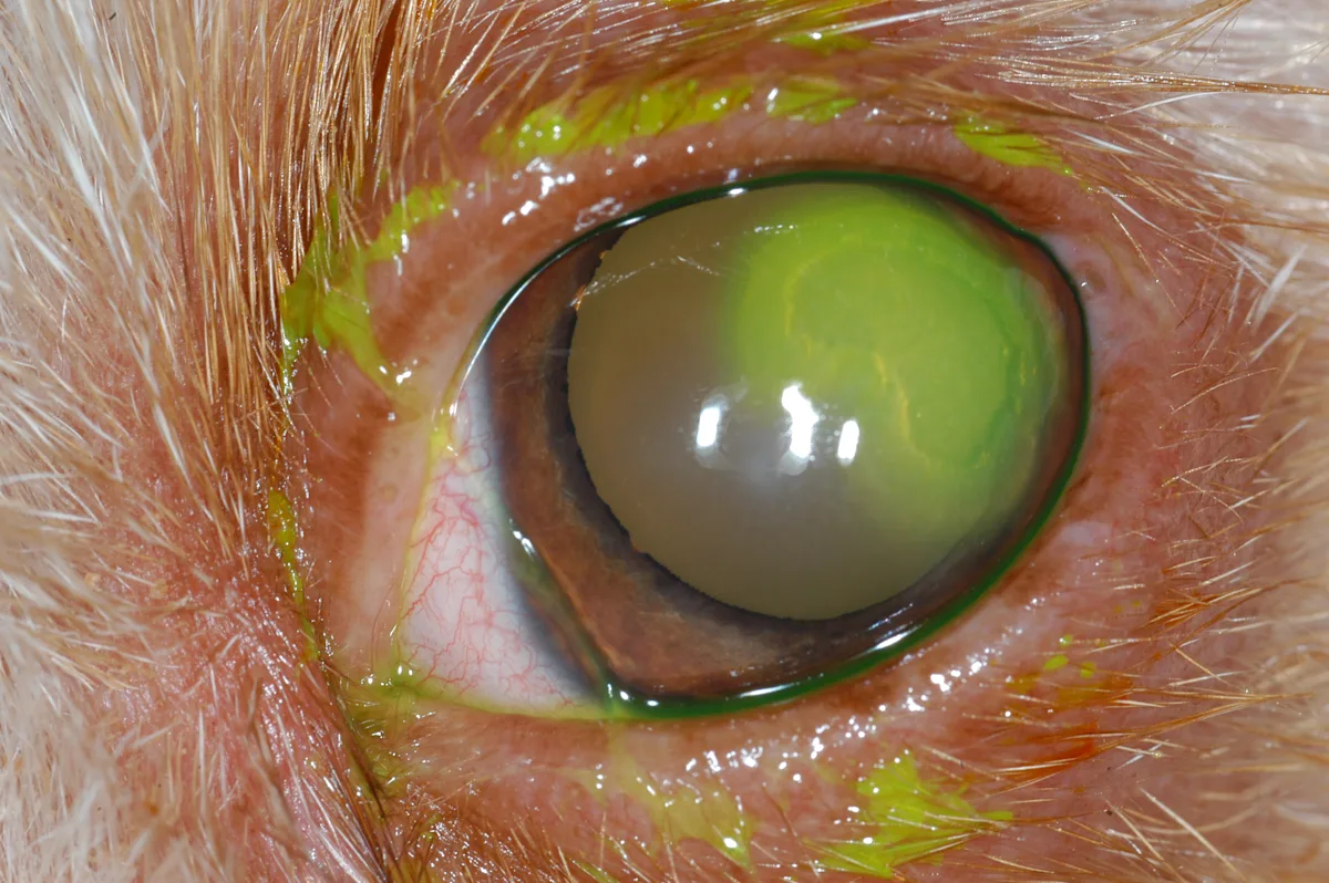
Explore More About Glaucoma & Other Ocular Diseases
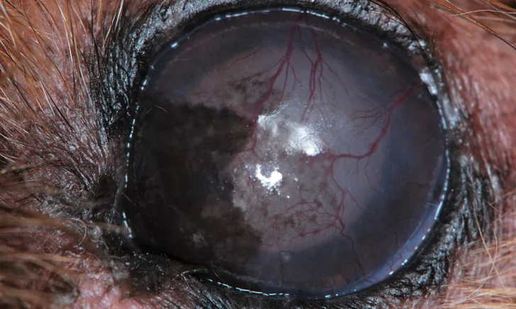
Ocular abnormalities may reflect serious underlying systemic disease. Know what to look for when performing an ophthalmologic examination.
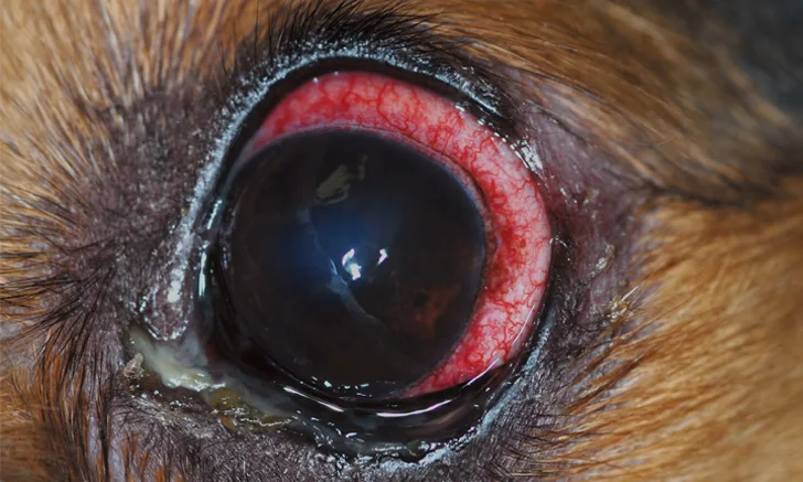
Common underlying causes of conjunctivitis vary greatly between dogs and cats, requiring different treatment approaches for each. Identify the best drug for every patient with the help of this practical reference guide.
Test Yourself
Clinical Challenge
Poll
Quiz
Clinical Image
Fact or Fib?
Clinical Scenario

Listen In
Clinician's Brief Podcast
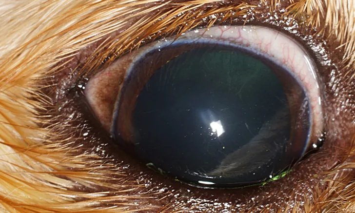
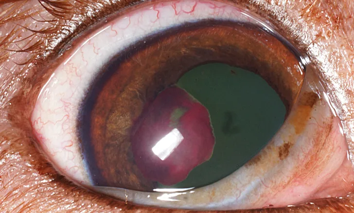
Listen in as ophthalmology expert Dr. Haeussler discusses the epiphora-related conditions that should be addressed and which can be lived with. Plus, he shares the 3 main ocular signs he suggests clients keep an eye out for.
Dr. Haeussler shares some fundamental ways to improve eye care at the general practice level, including what you need to be a better ocular photographer. He explains the advantages of going slow and encourages all of us to look at more eyes—the more you look, the more you’ll see.
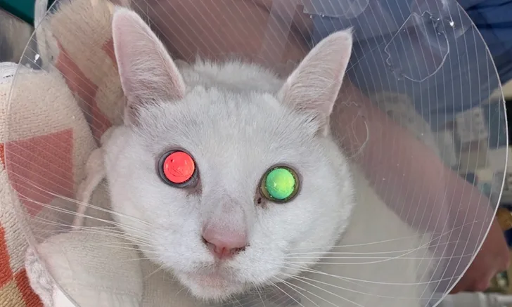
Dr. Brown shares what you should look for and how to look for it in these cases. He highlights some of the biggest differences between cats and dogs and why antihypertensive treatment should be initiated gradually. Don’t miss Dr. Brown’s tips for using your smartphone to capture ocular images.
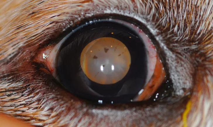
Dr. Lewin brings us all up to speed on golden retriever pigmentary uveitis, a prevalent hereditary ocular condition. He shares the most important examination findings and offers tips on practical eye examination techniques.
Preview Plumb's
See how Plumb's can answer your treatment FAQs for GI diseases.

Continuing Education
Earn CE credit with these free courses.
2-Minute Takeaways
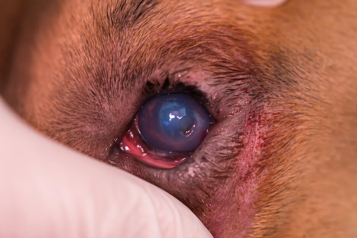
Congenital ocular malformations are rare, but it is important to recognize when they are present. Do you know which are most common in dogs and cats?
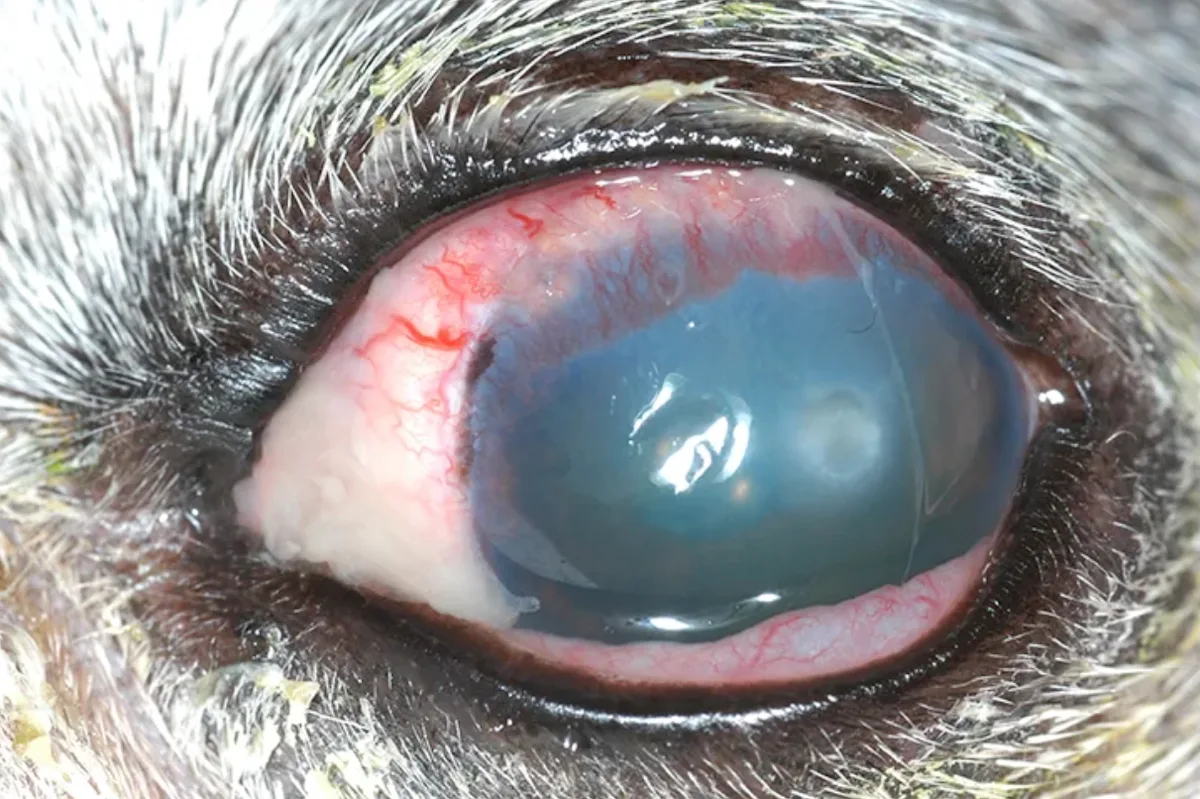
Infectious keratitis requires early and aggressive treatment to preserve ocular function. Ultraviolet light therapy may be an option to reduce bacterial growth while awaiting culture results.
Bacterial keratitis in cats is relatively uncommon but clinically important. Use this study to formulate an appropriate antibiotic plan for affected patients.
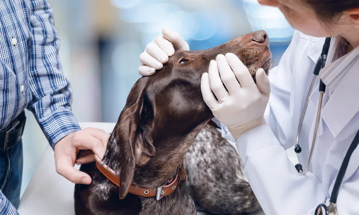
Neurogenic keratoconjunctivitis sicca (nKCS) has been commonly linked to trauma and otitis media. Discover the common characteristics of nKCS that developed in 29 dogs following treatment with a long-lasting otic medication.
Recommended Reading

Transpalpebral enucleation minimizes the risk for residual conjunctival tissues, making it the preferred approach when ocular neoplasia or infection is present. Feel confident in your technique with expert tips and procedural pictures found in this step-by-step guide.
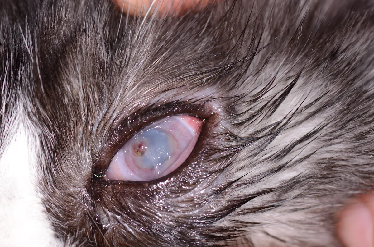
This case-based quiz tests your knowledge of ophthalmic medication selection in dogs and cats, with a focus on solutions vs ointments.

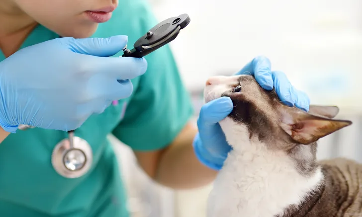
A good ocular examination starts with the right equipment and the right technique. Follow this step-by-step guide to performing direct ophthalmoscopy.
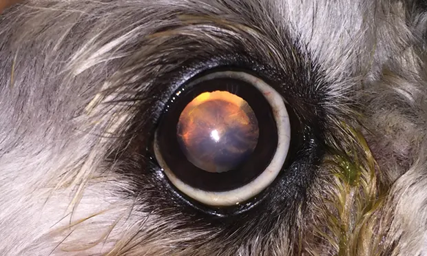
Follow a step-by-step guide to distinguish nuclear sclerosis from cataracts, and identify the underlying causes of each.