Top 5 Corneal Diseases in Aging Dogs
Kimberly Crane, DVM, Bliss Animal Eye Care, Medford, Oregon
Braidee Foote, DVM, DACVO, University of Tennessee
The cornea becomes increasingly susceptible to degenerative and inflammatory diseases as dogs age. Early recognition and appropriate treatment can preserve vision and ocular comfort in geriatric dogs.
Following are the top corneal diseases that affect geriatric dogs according to the authors.
1. Spontaneous Chronic Corneal Epithelial Defect
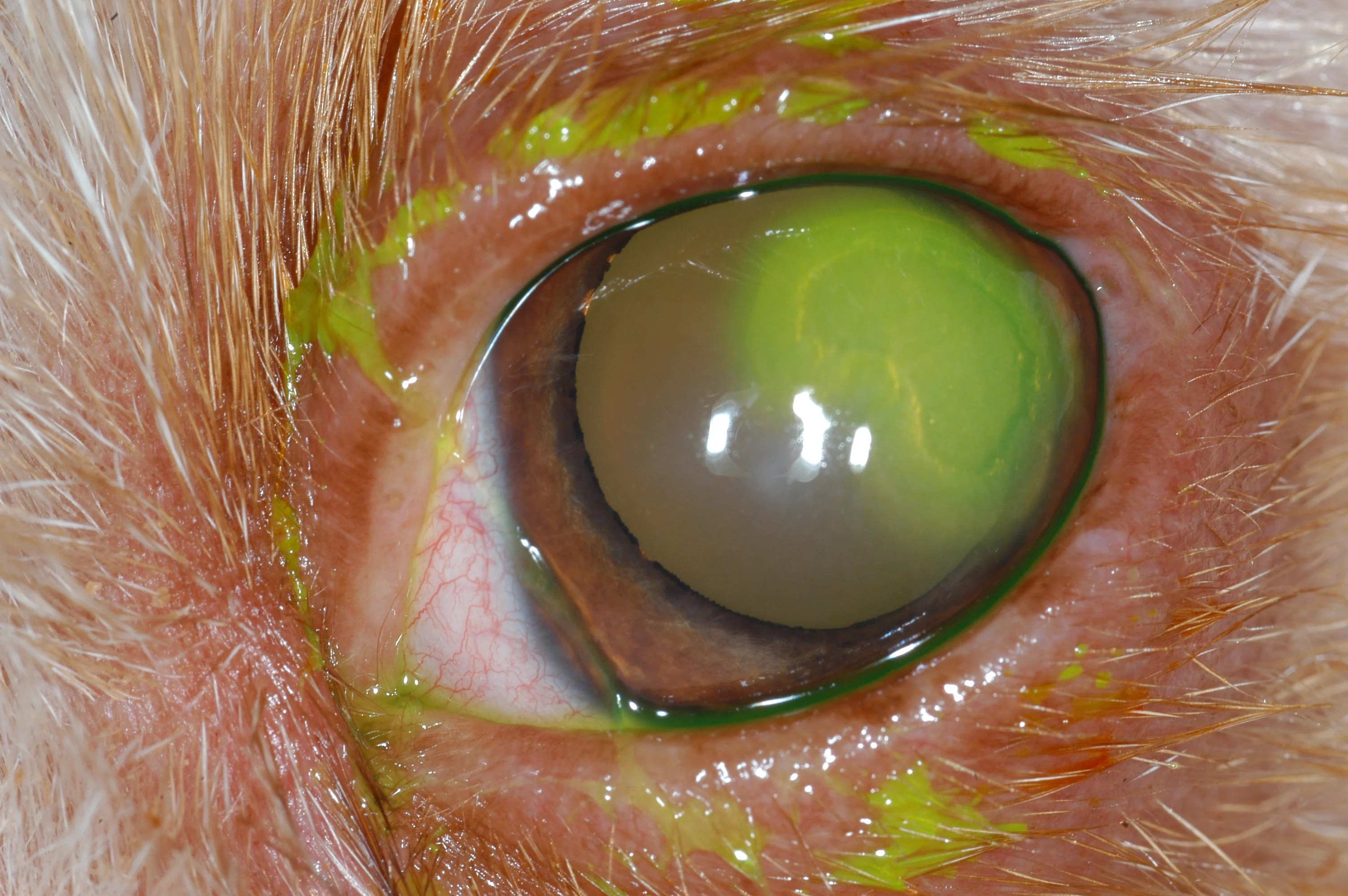
Spontaneous chronic corneal epithelial defects (SCCEDs; ie, indolent ulcers) are superficial, chronic, noninfected ulcers refractory to healing due to failure of the corneal epithelium to adhere to the underlying stroma.1-4 SCCEDs are common in older dogs of any breed, have an average age of onset of 8 to 9 years, and are particularly prevalent in boxers.2,3 Other breeds commonly affected include Staffordshire bull terriers, French bulldogs, Pembroke Welsh corgis, American bulldogs, Boston terriers, and English bulldogs. Loose corneal epithelium surrounds the ulcer and allows fluorescein stain to bleed under the edges, creating indistinct margins (Figure 1). Accurate diagnosis is critical, as treatment differs from other nonhealing ulcers.
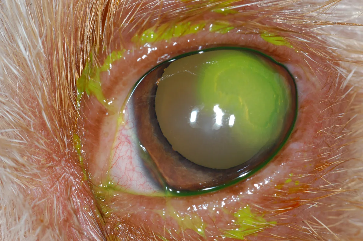
FIGURE 1 A spontaneous chronic corneal epithelial defect with the pathognomonic appearance of loose epithelial margins and a halo effect of fluorescein stain extending beyond the ulcer edges
Treatment of SCCEDs involves epithelial debridement with a cotton-tipped applicator and/or an anterior stromal modifying procedure (eg, diamond burr debridement, grid keratectomy) paired with routine ulcer management to improve success rates.2-4 Routine ulcer management includes topical broad-spectrum antibiotics administered 3 to 4 times per day, topical cycloplegics (eg, atropine) and/or oral analgesia (eg, NSAIDs), and an Elizabethan collar. A bandage contact lens may be placed after debridement to potentially speed healing and improve comfort.5
2. Corneal Degeneration
Corneal degeneration involves deposition of calcium, cholesterol, and/or other lipids within the cornea in association with inflammation.1-3 These corneal deposits occur spontaneously or secondary to systemic and ocular conditions (eg, kidney disease, hypertriglyceridemia, hyperadrenocorticism, dry eye, episcleritis, pannus).2,3 Calcium deposits are most common in older dogs, with an average age of onset of 13 years.6 Secondary inflammation, vascularization, pigmentation, and subsequent corneal ulceration can also occur. Deposits appear as asymmetric, sparkly silver to white opacities that can be unilateral or bilateral and are often axial in location (Figure 2). Diagnosis is based on appearance after a complete ocular examination.6 The underlying cause may be identifiable with full, fasted blood work that includes endocrine testing.
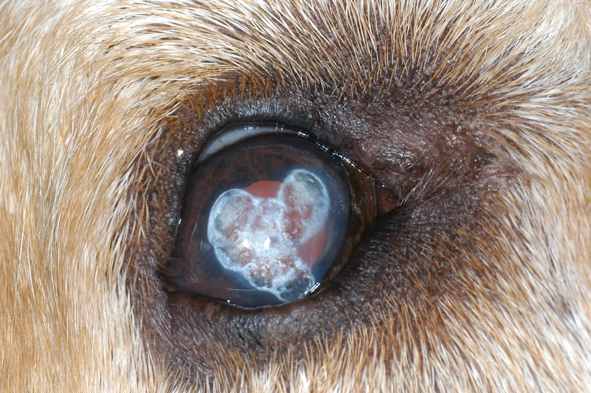
FIGURE 2 Appearance of extensive corneal degeneration with axial, crystalline, hazy, white opacities; evidence of keratitis can be seen as corneal vascularization extending from the medial and dorsal limbus to the axial deposits.
Treatment of corneal degeneration is directed at the underlying cause, if identifiable, and prevention of ulceration. Nonprogressive, mild deposits do not warrant treatment. Progressive lesions can be treated with topical 1% edetate disodium (disodium EDTA) 2 to 4 times per day to decrease progression of calcium deposition and secondary ulceration. Secondary ulceration should be treated routinely, with addition of disodium EDTA2,7; however, caution is needed, as these secondary ulcerations can be superficial or deep. Debridement of superficial ulcers and associated degenerative crystalline foci with a diamond burr can successfully remove and smooth deposits, allowing the cornea to heal; however, deeper stromal ulcerations should be referred for corneal stabilization surgery because of risk for corneal perforation.7
3. Keratoconjunctivitis Sicca
Keratoconjunctivitis sicca (ie, quantitative dry eye) occurs as a result of a deficiency in the precorneal tear film. The prevalence of dry eye increases in dogs >10 years of age, likely due to age-related lacrimal gland atrophy, but immune-mediated destruction is also common.8-11 Additional etiologies are metabolic (eg, diabetes mellitus, hypothyroidism), infectious, drug-induced, neurogenic, radiation-induced, iatrogenic, and idiopathic.2,8 Many breeds are predisposed, including American cocker spaniels, Cavalier King Charles spaniels, West Highland white terriers, and brachycephalic breeds.9-11 Clinical signs include mucopurulent discharge, corneal vascularization and pigmentation, conjunctival hyperemia, and possible subsequent corneal ulceration (Figure 3). These signs can be insidious and bilateral in older dogs and lead to decreased vision if left undiagnosed and untreated. Diagnosis is established with a Schirmer tear test <10 mm/minute or <15 mm/minute with associated clinical signs.8,11
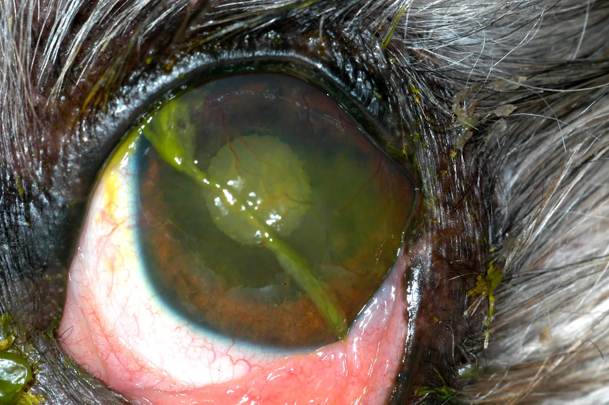
FIGURE 3 Chronic keratoconjunctivitis sicca causing keratitis (ie, corneal vascularization), fibrosis, and conjunctival hyperemia; mucoid discharge is also adhered to the cornea and periocular fur.
Treatment of keratoconjunctivitis sicca is often lifelong and involves tear stimulation (eg, 0.2%-2% cyclosporine A, 0.03%-1% tacrolimus 2-3 times per day) and corneal lubrication with gel or ointment.8,9,11 Surgical transposition of the parotid salivary gland duct can lubricate the ocular surface in severe, refractory cases.9
4. Corneal Endothelial Degeneration
Corneal endothelial degeneration (CED; ie, corneal endothelial dystrophy) is characterized by progressive loss of corneal endothelial cells, resulting in bilateral, advanced, and permanent corneal edema. Advanced corneal edema significantly alters vision and can lead to bulla formation, rupture, and ulceration (Figure 4).1-3,12 Several breeds (eg, Boston terriers, dachshunds, Chihuahuas) are thought to be predisposed to CED.2,3,12 Corneal edema begins at an average 12 years of age with an asymmetric, temporal, light-blue haze that progresses to bilateral diffuse edema.1,2,12 Diagnosis is based on signalment and exclusion of other causes of bilateral diffuse corneal edema (eg, anterior uveitis, anterior lens luxation, endotheliitis, glaucoma).
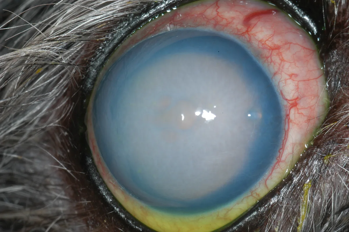
FIGURE 4 Severe, diffuse corneal edema with a central large bulla caused by CED; left untreated, the bulla can rupture and lead to corneal ulceration.
Application of topical 5% sodium chloride 3 to 4 times per day can reduce epithelial bullae but cannot reverse stromal edema.3,12,13 Surgical options include thermokeratoplasty or superficial keratectomy and conjunctival advancement hood flap.14,15 Descemet’s stripping endothelial keratoplasty, a type of corneal transplant, has been evaluated in dogs but remains challenging because of the need for fresh donor tissue.16
5. Pigmentary Keratitis
Pigmentary keratitis (ie, corneal melanosis) develops secondary to chronic inflammation, resulting in a progressive dark pigment that spreads across the cornea and affects the vision (Figure 5). This condition can occur in any breed but is most common in brachycephalic breeds (eg, pugs, Boston terriers, bulldogs).3 One study reported a 10% increased risk for corneal pigmentation per year of age in brachycephalic dogs.17 Underlying causes (eg, mechanical abrasion, tear film disorders, immune-mediated keratitis [eg, pannus], chronic corneal ulcers [infected or indolent]) should be identified and treated when pigmentation is diagnosed.1,18
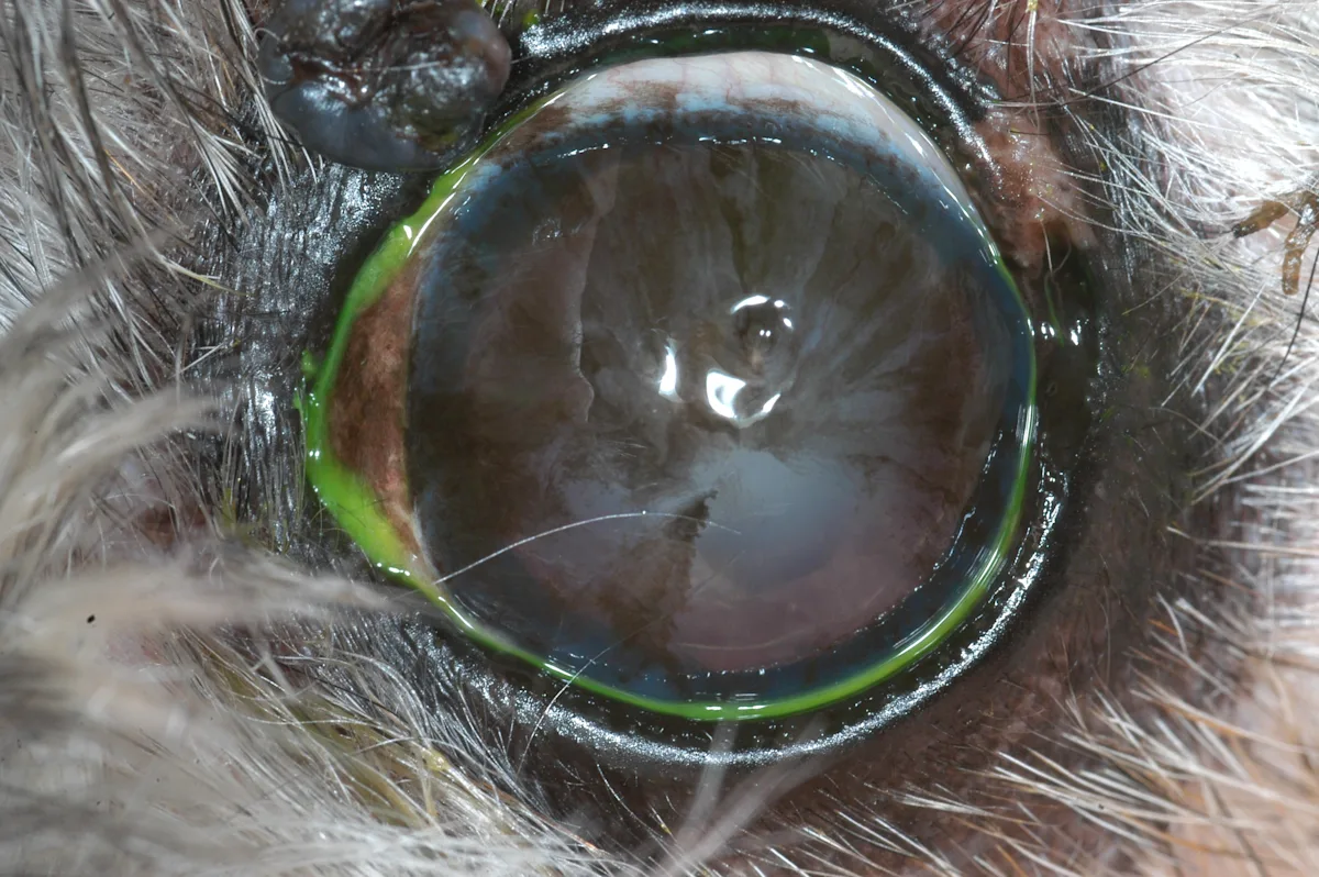
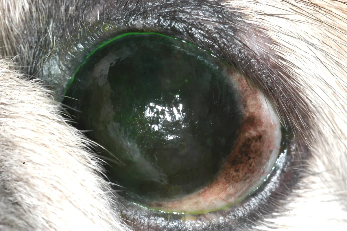
FIGURE 5 Pigmentary keratitis leading to wispy, superficial corneal pigment affecting 75% of the corneal surface after healing a centrally located infected corneal ulcer seen in a shih tzu, which is a geriatric predisposed breed (left). Severe pigmentary keratitis completely occluding vision due to chronic untreated dry eye disease seen in a Pekingese, which is a geriatric predisposed breed (right).
Nonspecific treatments focus on decreasing inflammation and halting the spread of pigment to maintain vision. Topical cyclosporine A, tacrolimus, and prednisolone acetate have been used with varying degrees of success.1 Of these treatments, tacrolimus is most promising for reducing existing pigmentation rather than simply slowing progression.19 Superficial keratectomy, radiation therapy, or cryosurgery can be performed to reduce blinding pigmentation in severe cases, but recurrence of pigment is inevitable.20
Conclusion
Accurate diagnosis, early detection, and targeted management of these common corneal pathologies can improve long-term comfort, vision, and quality of life in aging dogs.