Cardiac Library
Cardiac Examination
Approaching the cardiac patient requires basic knowledge of what to look for in the patient history, cardiac auscultation technique, diagnostic imaging of the heart, and cardiac electrophysiology. In this section, clinicians can learn to hone these critical skills to get the most from their cardiac exam.
The best stethoscope is not necessarily the most expensive. One expert shares her tips on selecting and using this essential diagnostic tool.
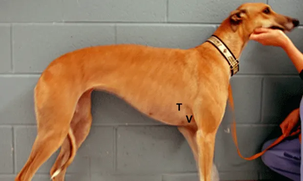
An accurate physical examination and clinical history allow a clinician to focus the diagnostic workup so that superfluous diagnostic tests can be avoided.
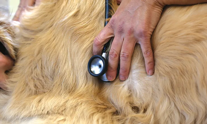
There are 12 components of a basic cardiology examination. Can you name them all?
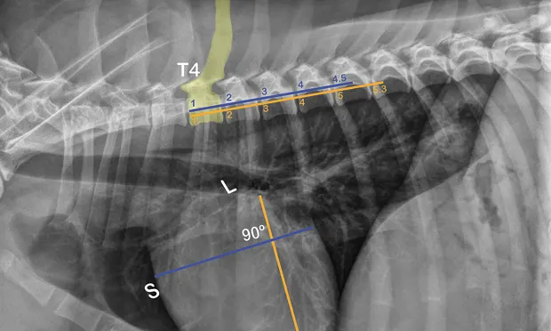
This cardiologist guides you through calculating a vertebral heart scale, its uses, and interpreting findings.

Vertebral heart scale (VHS) is an objective radiographic measurement that can be useful when cardiac disease is suspected. Review how to calculate VHS in cats.

Vertebral left atrial size (VLAS) is an objective radiographic measurement that can help determine whether left atrial enlargement is present with suspected or diagnosed myxomatous mitral valve disease. Review how to calculate VLAS in dogs.
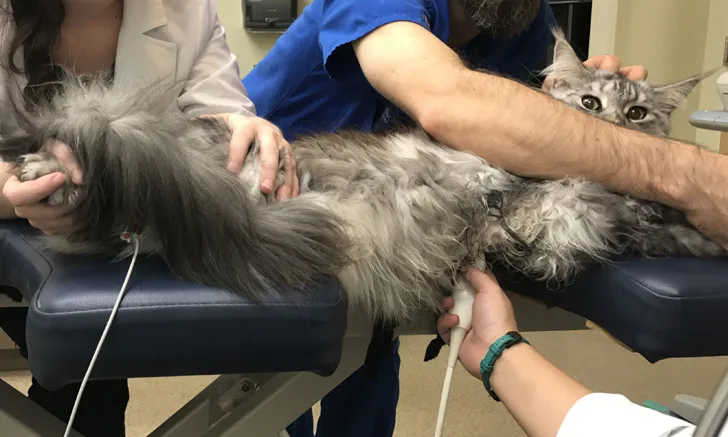
Learn key echocardiography views you can put to use in your practice today.
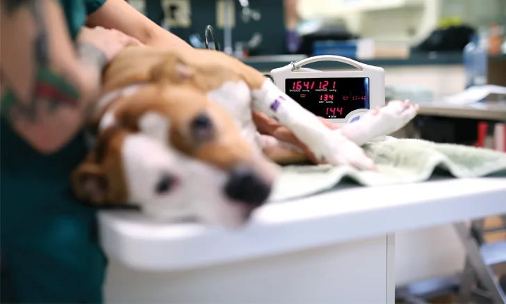
Blood pressure measurement is a simple diagnostic procedure that provides a wealth of information. Sharpen your skills with this comprehensive guide to obtaining accurate readings.
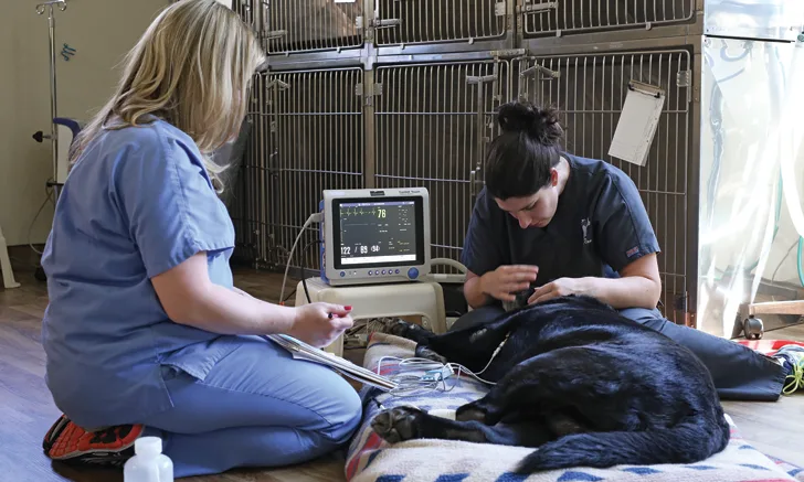
This step-by-step guide provides tips for obtaining an accurate diagnostic ECG on every patient, every time.

Confident ECG interpretation starts with a systematic approach. Review waveforms, intervals, heart rate calculation, and rhythm identification in this practical guide for small animal practitioners.
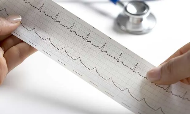
How do you interpret ECGs with confidence? This first installment of a two-part series outlines ECG interpretation.
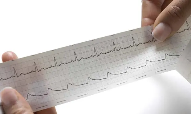
This is the second installment of a two-part series outlining ECG interpretation and addresses bradyarrhythmias and tachyarrhythmias.
Keep Your Finger On the Pulse
From podcasts with expert guests to quizzes to test your knowledge, dive deeper into cardiac care with our Cardiac Library.