The Basic Cardiology Examination
Wendy W. Mandese, DVM, University of Florida
Amara H. Estrada, DVM, DACVIM (Cardiology), University of Florida
For a comprehensive outline of cardiology history & diagnostics, see Clinical Cardiology History & Diagnostics by Drs. Mandese and Estrada.
A thorough physical examination is valuable for diagnosing heart disease and should include extensive examination of all body systems.
General Appearance
Weight loss is common in patients with advanced cardiac disease. In one study, >50% of dogs with dilated cardiomyopathy experienced cardiac cachexia (ie, loss of lean body mass).1 Low body weight and failure to grow normally can also be signs of congenital cardiac disease in pediatric patients.
Ocular Examination
Enlarged retinal vessels, retinal hemorrhage, or retinal detachment often indicates systemic hypertension, which can be primary (ie, idiopathic) or secondary to conditions such as renal or cardiac disease, hyperadrenocorticism, diabetes mellitus, pre-eclampsia, and hyperthyroidism. In patients with inconclusive laboratory results, echocardiography can be used to identify underlying heart conditions that cause hypertension (eg, hypertrophic cardiomyopathy in cats, myxomatous mitral valve disease and cardiac overload in dogs). Cardiac changes secondary to hypertension include myxomatous mitral valve disease, left atrial enlargement, or left ventricular hypertrophy.2
Oral Examination
Pale mucous membranes can indicate shock or anemia, and hyperemic or injected mucous membranes can indicate infection or polycythemia. Cyanosis occurs primarily in patients with cardiac defects that result in right-to-left shunting, such as reverse patent ductus arteriosus, atrial septal defect, and ventricular septal defect. Other health conditions, such as severe hypothermia or severe respiratory disease, can also cause cyanosis. In differential cyanosis, the lower extremities and vulva/prepuce appear cyanotic but the upper extremities and oral mucous membranes are pink and well oxygenated.3
Several congenital cardiac defects are associated with differential cyanosis, but it is most commonly seen with a reverse patent ductus arteriosus. Periodontal disease should be assessed and noted, as secondary cardiac complications (eg, endocarditis) are possible if severe periodontal disease is not addressed.4
Tracheal Palpation
Tracheal palpation should be performed on all coughing dogs. Small-breed dogs more prone to myxomatous mitral valve disease are also more prone to a collapsing trachea. In both of these conditions, cough can occur during exercise or excitement5 and may be caused by tracheal disease, even if a murmur is present. Coughing can be heard with tracheitis or tracheobronchitis and can be caused by bacterial or viral infection or environmental allergens.
Respiratory Rate/Effort
Respiration should be evaluated when the patient is calm. Clients can be trained to take respiratory rates at home, especially while the patient is sleeping. Several phone apps are available to make it easy for clients to obtain accurate readings. One study suggested that most dogs and cats without cardiac disease or with well controlled heart disease will have a resting respiratory rate of <30 breaths/min at home.6 Increased respiratory effort can indicate upper airway disease (effort occurs during inspiration) or lower airway disease (effort occurs during expiration).
Lung Sounds
Abnormal lung sounds are most common in patients with primary respiratory disease. Lung sounds also may be abnormal in patients with secondary respiratory conditions such as pulmonary edema and pleural effusion. Auscultation of the lungs is not a sensitive means of detecting pulmonary edema or pleural effusion in dogs and cats, and many patients have pulmonary edema with no auscultatory abnormalities other than increased bronchovesicular sounds.7 Patients with severe pulmonary edema resulting in free fluid in the airways are more likely to have audible crackles; crackles can also be auscultated in patients with pulmonary hypertension, bronchitis, and pneumonia. Muffled lung sounds can indicate pleural effusion. Wheezes are associated with allergic airway disease, bronchitis, and collapsing trachea.
Heart Rate
Stress and anxiety associated with the veterinary environment can markedly increase a patient’s heart rate. Waiting for a patient’s initial arrival excitement to subside and asking the client to be present during examination can help the clinician obtain a normal heart rate. The client can also be asked to obtain the patient’s resting heart rate at home. If an arrhythmia is present, the type (eg, tachyarrhythmia, bradyarrhythmia) should be noted and the nature characterized.
Pulse
Pulse pressure can be decreased or increased or have an altered configuration. Decreased pulse pressure may be seen in patients with dilated cardiomyopathy, aortic or pulmonic stenosis, heart failure, hypovolemia, or shock. Increased pulse pressure can occur because of excitement and/or pain or hypertrophic cardiomyopathy.8 Dogs with aortic regurgitation commonly have a bounding pulse. Bounding pulses can also be felt in patients with patent ductus arteriosus, severe bradycardia, hyperthyroidism, fever, or anemia.
Alterations in pulse conformation also may occur. Dogs with severe subaortic stenosis can have a weak pulse or a pulse pressure that increases more slowly and peaks later during systole (ie, pulsus parvus et tardus). Conversely, dogs with mitral regurgitation commonly have a brisk pulse that rises more rapidly in systole and lasts a shorter time. Other pulse abnormalities include pulsus paradoxus and pulse deficits. Pulsus paradoxus is an increase in pulse pressure on expiration and a decrease on inspiration. This occurs normally but is exaggerated in cardiac tamponade. Pulse deficits can occur with cardiac tachyarrhythmias in which beats occur so rapidly that the ventricle does not have time to fill with an adequate amount of blood before ejection (eg, fast atrial fibrillation, ventricular premature beats).7 Pulse should always be monitored while performing cardiac auscultation to detect pulse deficits.
Blood Pressure
Obtaining a systolic blood pressure using a Doppler is a relatively simple procedure. To obtain the most accurate reading, blood pressure should be measured in a quiet, calm environment with the client present, if possible. Proper technique, including appropriate cuff width (ie, 40% of the circumference of the limb or tail) is integral to obtaining an accurate measurement. A minimum of 3 measurements should be obtained, and the variability between measurements should be <20%.9
Heart Sounds
The first heart sound (S1) is produced by closing of the mitral and tricuspid valves. S1 is loudest over the mitral valve area and is louder, longer, and lower pitched than the second heart sound (S2). S2 is produced by closing of the pulmonic and aortic valves. S2 is shorter and higher pitched than S1.
The third heart sound (S3) is not usually heard during auscultation of healthy small animals, and its presence indicates myocardial failure. The sound is generated during the period of rapid filling in early diastole when the ventricles suddenly resist expansion.
The fourth heart sound (S4) originates from the vibration generated by cardiac structures when the atria contract. It can be a normal finding in giant-breed dogs and large animals or be associated with advanced hypertrophic cardiomyopathy. The presence of an S3 or S4 is referred to as a diastolic gallop. When a diastolic gallop is present, further evaluation is warranted.
Systolic clicks are found in dogs with chronic valvular disease and originate from vibrations that occur when chordae tendineae and the mitral leaflets suddenly resist further stretching and protrude into the left atrium.10 Muffled heart sounds may indicate the presence of pericardial or pleural effusion.
Arrhythmias
Sinus arrhythmia is common in dogs, especially in patients who are relaxed and have a lower heart rate. The heart rhythm is faster on inspiration and slower on expiration. When the patient becomes more excited or active, the sinus arrhythmia is often no longer heard. Sinus arrhythmia does not always correspond to respiration and can occur with other causes of increased vagal tone (eg, GI disease).11 Pulses should be palpated in conjunction with auscultation to determine if pulse deficits are present. Sinus arrhythmia was previously thought to be uncommon in cats, but a 2009 study determined that relaxed cats in their home environment can have frequent sinus arrhythmia.12
Common abnormal rhythms include:
Premature beats with pulse deficits: Associated with atrial and ventricular premature complexes. Can occur in bursts or be sustained
Irregularly irregular rhythm: Associated with atrial fibrillation. Has been described as “shoes in a dryer”
Slow arrhythmia with intermittent pauses: Heard in patients with AV block or sinus arrhythmia
Persistent tachycardia or persistent bradycardia: Clients should be informed that an abnormality in heart rate and/or rhythm requires additional testing to determine the cause.
Murmurs: Caused by turbulence disturbing the normal laminar flow of blood, which most often is caused by dysfunctional valves or septal defects. Can also have physiologic causes because of patient size, athletic ability, or underlying noncardiac disease processes (eg, fever, anemia)13
Characterization of murmurs is based on several criteria:
Timing in the cycle: A systolic murmur occurs between S1 and S2 and is common in small animals. A diastolic murmur occurs between S2 of one beat and S1 of the following beat and is uncommon in small animals. A continuous murmur can be heard throughout the cardiac cycle (Table 1).
Table 1: Common Pathologic Causes of Murmurs Based on Timing7
Location: Location or point of maximal intensity (PMI) refers to the valve area at which the murmur is heard loudest. Recognition of the PMI can help identify the specific cardiac abnormality (Table 2; Figures 1-5).
Table 2: Localization of Murmurs in Common Cardiac Lesions7

FIGURE 1 Position of cardiac apex and base in a left canine thorax. Illustrations by Ally Mandese and used with permission
Intensity
Murmurs are graded on a scale of 1 to 6. This scale can be subjective and may vary between observers.
Grade 1/6: Softest murmur audible. May only be heard in a quiet room after listening for several minutes. May not be audible to all observers, and may be transient
Grade 2/6: Soft but more easily heard. Often focal over one valve only
Grade 3/6: Prominent and easily heard. May radiate to other areas
Grade 4/6: Loud and radiates widely. Not accompanied by a palpable thrill
Grade 5/6: Loud and accompanied by a palpable thrill
Grade 6/6: Loud, accompanied by a palpable thrill, and can be heard with the stethoscope barely touching the thorax
Innocent murmurs are soft, systolic, heard best at the mitral or aortic valves, and often low-grade (grade 1). They do not radiate. They are often auscultated in young puppies and kittens and usually disappear by 3 to 4 months of age.
Physiologic murmurs are soft and usually of low intensity (grade 1-2). PMI is at the heart base in the area of the outflow tracts (aortic and pulmonic area). These murmurs typically resolve with resolution of the underlying disease. They may also be heard in otherwise healthy animals that are deep chested and/or athletic. They do not indicate any underlying cardiac disorder that can be identified by chest radiography or echocardiography and are common in animals with anemia and occurs as a result of changes in blood viscosity.
Character: Most murmurs have characteristic sounds on auscultation:7
Harsh or regurgitant: Mimicked by placing the back of the tongue close to the roof of the mouth and blowing out forcefully. Heard in patients with ventricular septal defects and atrioventricular valve insufficiency
Blowing: Mimicked by blowing air with moderate force through slightly parted lips. Often heard in patients with aortic or pulmonic insufficiency
Machinery: Sounds like the wind blowing through a tunnel. Most often heard in patients with patent ductus arteriosus
Systolic clicks: High frequency sounds heard over the left apex that may indicate mitral valve disease
Crescendo-decrescendo: An ejection murmur most common in patients with atrial septal defects and aortic or pulmonic stenosis
Heart murmurs are present in approximately one-third of apparently healthy adult cats.14-16 The intensity of these murmurs can vary with sympathetic tone and usually increases as heart rate increases. The murmur can disappear entirely when sympathetic stimulation abates and heart rate slows. A murmur may be audible on initial auscultation and may soften or disappear as the patient relaxes during the examination.
Hypertrophic cardiomyopathy and systolic anterior motion of the mitral valve are the most common diagnoses in cats with murmurs caused by heart disease.14-16 Because benign murmurs and murmurs caused by cardiac disease are dynamic and audibly indistinguishable, further evaluation is warranted when a murmur is detected.17
Abdominal Palpation
Ascites and/or liver enlargement may be present in patients with right-sided heart failure.
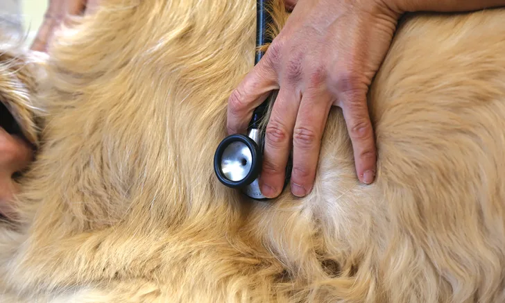
Step by Step: Cardiac Auscultation
Cardiac auscultation is best performed in a quiet room with a standing patient. In anxious dogs, panting sounds can be mistaken for murmurs. Manually close the dog’s mouth for a short time during auscultation and give panting breaks as needed. Purring in cats can also be mistaken for a murmur. Distract the cat with a toy or change the position or location of the patient to stop purring.
Step 1
Place the stethoscope over the left cardiac apex (location of ventricles) and base (location of pulmonary and aortic outflow tracts), right cardiac apex and base, the length of the sternum, and the thoracic inlet.
Step 2
Auscultate each heart valve at its PMI (Table 3).
Table 3: Stethoscope Placement For Cardiac Examination
Conclusion
Cardiac abnormalities can be identified during a basic cardiac examination that includes obtaining a complete history and performing a thorough physical examination and simple diagnostics. Creating a list of differential diagnoses based on the information gathered during the cardiac examination can help the clinician make informed decisions about the appropriate next diagnostic or therapeutic step.