Vertebral Heart Scale
Stacey Fox-Alvarez, DVM, MPH, DACVIM (Oncology), University of Florida, Community Care Veterinary Specialists, Gainesville, Florida
Amara H. Estrada, DVM, DACVIM (Cardiology), University of Florida
How do I calculate a vertebral heart scale (VHS)?
The vertebral heart scale system (ie, vertebral heart size) was developed as a means to objectively evaluate cardiac size among dogs of different breeds and thoracic conformations.
Uses of Vertebral Heart Scale
The VHS was established to create a more objective way of diagnosing cardiomegaly via thoracic radiography. Individual dogs can have values that fall outside of the normal range without cardiac disease, so it should not be used as the only means of diagnosing cardiac disease in any given patient.
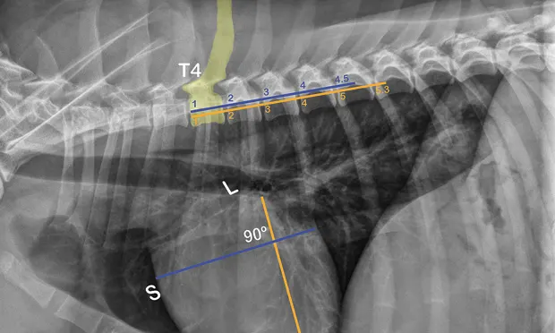
In addition to a thorough history and physical examination, VHS can be used to raise clinical suspicion of heart disease. VHS is useful for monitoring a patient in which heart disease is suspected (Figure 1), or in tracking changes in progressive cardiac disease over time.

Illustration of VHS calculation in a normal dog. On a lateral radiograph, a line is drawn from the carina to the most ventral aspect of the heart. This line is the “L” or long axis line. A line is drawn perpendicular to the long axis at the widest portion of the heart, extending to the cranial and caudal borders. This is the short axis or “S” line. These lines are transposed using calipers extending along the spine from the cranial aspect of T4. The number of vertebrae traversed (rounded up to the nearest tenth) are added together for the VHS.
VHS has been shown to correlate with other means of measuring cardiomegaly (ie, ECG, echocardiography) in dogs with progressively increasing heart size, and it is considered by some to be the gold standard in determining cardiomegaly in dogs.1 The authors believe many dogs with presumptive MVD can be managed effectively by use of a thorough physical examination and basic diagnostics, such as a VHS and measurement of blood pressure. As guidelines for the treatment of heart disease in veterinary medicine evolve, it will become increasingly important to accurately stage patients with these conditions to determine when medical treatment is indicated. VHS calculation can be an important component to the staging of cardiac disease.
Pimobendan and ACE inhibitor treatment for Doberman pinschers with preclinical dilated cardiomyopathy (DCM) have both been shown to delay onset of clinical signs2,3 such as sudden death or congestive heart failure (CHF) and should be considered when imaging yields evidence of preclinical disease (ie, left ventricular enlargement, decreased contractility identified on echocardiography). Results are mixed when evaluating the use of ACE inhibitors in preclinical dogs with stage B2 MVD.4,5
ACE inhibitors are currently recommended for these patients by some specialists, as well as the majority of the members of the ACVIM consensus panel for MVD.6 As more studies evaluate the use of medical intervention to delay the onset of CHF in dogs with preclinical cardiac disease, treatment recommendations will continue to change.
Quiz Yourself on Measuring Heart Scale!
Already a pro at measuring VHS? Take our VHS quiz here!
Serial measurements and rate of change of VHS have been demonstrated to be predictive of the onset of congestive heart failure in Cavalier King Charles spaniels with mitral valve regurgitation.7,8 Several studies in this breed have shown that the rate of change of VHS on serial radiography (ie, every 6-12 months) remains steady up until 6-12 months prior to onset of CHF, when it increases at a much faster rate.7,8
The ACVIM consensus statement on degenerative MVD recommends baseline thoracic radiography for dogs with a new murmur, then annually thereafter.6 For practitioners adhering to these guidelines, annual calculation of VHS and rate of change from previous imaging should become standard practice and may help identify those patients at higher risk of developing CHF in the coming year. Figure 2 illustrates annual radiographs taken in a patient with progressive cardiomegaly.

FIGURE 2A
Progressive cardiomegaly noted on annual evaluations in a dog with MVD.
The VHS has been shown to be useful when determining whether a cough is cardiogenic, pulmonary, or mixed in origin for dogs with MVD.9 One study found that a VHS >12.0 was suggestive for a cardiac component to cough in dogs with MVD.9 It is important to note that among these dogs, some were determined to have a cough of mixed cardiac and pulmonary origin.
VHS has also been shown to be useful when diagnosing CHF as a cause of dyspnea in cats, which is notoriously difficult because of the varied appearance of pulmonary edema in this species. One study found that a VHS >9.3 was highly sensitive for CHF as a cause of dyspnea and that cats with a VHS <8.0 were unlikely to have dyspnea secondary to cardiac disease.10 Unfortunately, the presence of pleural effusion, which is common in cats with heart failure, can obscure the cardiac apex. This makes the VHS calculation difficult or impossible to accurately determine in some animals.10
Related content: Vertebral Heart Scale Reference Intervals for Small Breed Dogs
Normal Values of Vertebral Heart Scale in Dogs
The short and long axis measurements can also be taken on ventrodorsal (VD) or dorsoventral (DV) projections, but this measurement is not as consistent between animals. Measurement from a lateral projection is recommended, if possible. The reference values for vertebral heart scale measured on VD and DV radiographs are 9.4-11.0 and 8.8-11.7, respectively, for dogs.11
The published range for a normal VHS is 9.2-10.3 on a lateral radiograph, with 10.5 suggested as the cutoff for clinical determination of cardiomegaly in adult dogs.11
The original guidelines for VHS were designed to be applicable across breeds and conformations, and most dog breeds studied individually fall within the normal range. However, several breeds have normal VHS values that would suggest cardiomegaly using the original scale.
Of the breeds evaluated to date, the boxer, bulldog (French and English), Boston terrier, Cavalier King Charles spaniel, Labrador retriever, pug, Pomeranian, and whippet have been found to have average values that are much higher than other dog breeds. New sets of normal values have been developed for use in these breeds12-14 (Table).
A normal range of 6.7-8.1 has been established for cats.15
Table: Normal Values for Select Dog Breeds
Step-by-Step: How to Calculate Vertebral Heart Scale (VHS)
What You Will Need
To calculate a VHS, a lateral thoracic radiograph in right or left lateral recumbency is needed. If using plain films, calipers or a ruler will be necessary to hold the measurements. If using digital films, calipers or electronic calipers can be used.
The best possible technique and positioning of the patient should be used to prevent rotation from affecting measurements and to make serial imaging comparisons more reliable.
Step 1
Using the calipers, measure the long axis of the heart from the carina to the apex of the heart at its most ventral point.
Step 2
Starting at the cranial aspect of the fourth thoracic vertebrae (T4), transfer this measurement (ie, heart length) to extend caudally along the thoracic vertebrae at mid-body position (Figure 1). Add the number of vertebrae this measurement traverses. Round to the nearest tenth of a whole number. This is measurement “L.”
Step 3
Measure the heart at its widest point (ie, the short axis) perpendicular to the line drawn for long axis.
Step 4
Starting at the same point on T4, transfer this measurement to extend caudally along the thoracic vertebrae at mid-body position and add the number of vertebrae it traverses. Round to the nearest tenth of a whole number. This is measurement “S.”
Step 5
To calculate the vertebral heart scale, add the “L” and “S” measurements together.
Author Insights
Right or left lateral recumbency is acceptable. If taking serial images, they should be done using the same side.
The long axis should be drawn from the level of the carina. The short axis should be perpendicular to the long axis.
Typically, a value of <8 for cats is used for convenience (normal, 6.7-8.1). A value >8 indicates cardiomegaly, which may indicate underlying pathology.
In cats, normal VHS does not rule out heart disease or dysfunction, as diseases that produce concentric hypertrophy (eg, hypertrophic cardiomyopathy) will not necessarily be accompanied by an enlarged cardiac silhouette.
Noncardiac Factors That Can Affect VHS
Interobserver variability is a known factor affecting consistent measurement of a VHS. One study showed an average difference between individuals of around 1 vertebrae when evaluating the same films.16 Ideally, the same veterinarian should calculate the VHS at all time points for an individual patient when tracking changes.
Abnormal vertebrae, such as hemi-vertebrae, are common in breeds such as Boston terriers and bulldogs. When present, these will impact the VHS. The study establishing the normal values for these breeds cited this as one of the reasons for their higher values.12
Pericardial effusion has been shown to cause an increased VHS result. In 1 study, the majority of dogs with pericardial effusion had a VHS >12.0.17 This should be a consideration for any dog with a generalized cardiomegaly on thoracic radiography. A thorough physical examination and history can help to rule this condition in or out as a cause for exercise intolerance, lethargy, collapse, ascites, or other similar signs.
Prolonged moderate-to-severe anemia in cats and dogs has been shown to cause compensatory left-sided cardiomegaly and an increase in the VHS measurement.18,19 In these patients, cardiomegaly is thought to occur secondary to volume overload via activation of the renin-angiotensin system, as well as through other neuroendocrine mediators.20 Initially, left atrial enlargement develops, which is followed by left ventricular hypertrophy. Cats can eventually develop CHF.18,19
Right vs left lateral recumbency may impact the VHS. Several studies have shown no difference between measurements, while others have shown that dogs in right lateral recumbency have higher VHS values (by approximately 0.3 vertebrae).1,4,21,22 This suggests that serial measurements of the same patient should be performed with consistent positioning; however, this may not be clinically significant.
Other Considerations
It is important to note that a normal heart size on thoracic radiography does not rule out cardiac disease or dysfunction. The VHS is a valuable tool to assess cardiac status and should be used in conjunction with other diagnostics (ie, physical examination, history, resting respiratory rate, ECG, NT-proBNP, echocardiography) to determine if treatment for cardiac disease is warranted or if further testing is needed based on clinical suspicion of cardiac involvement in disease.