Cardiac Library
Evaluation of the cardiovascular system involves more than simply listening through a stethoscope. Thorough evaluation also requires visual assessment of potential clinical signs, palpation to assess pulses and thrills, and clinical reasoning when evaluating patient history and signalment. This resource can aid clinicians seeking to elevate their knowledge of cardiac assessment, diagnosis, and management.




Relevant Content

Vertebral left atrial size (VLAS) is an objective radiographic measurement that can help determine whether left atrial enlargement is present with suspected or diagnosed myxomatous mitral valve disease. Review how to calculate VLAS in dogs.

There are 12 components of a basic cardiology examination. Can you name them all?
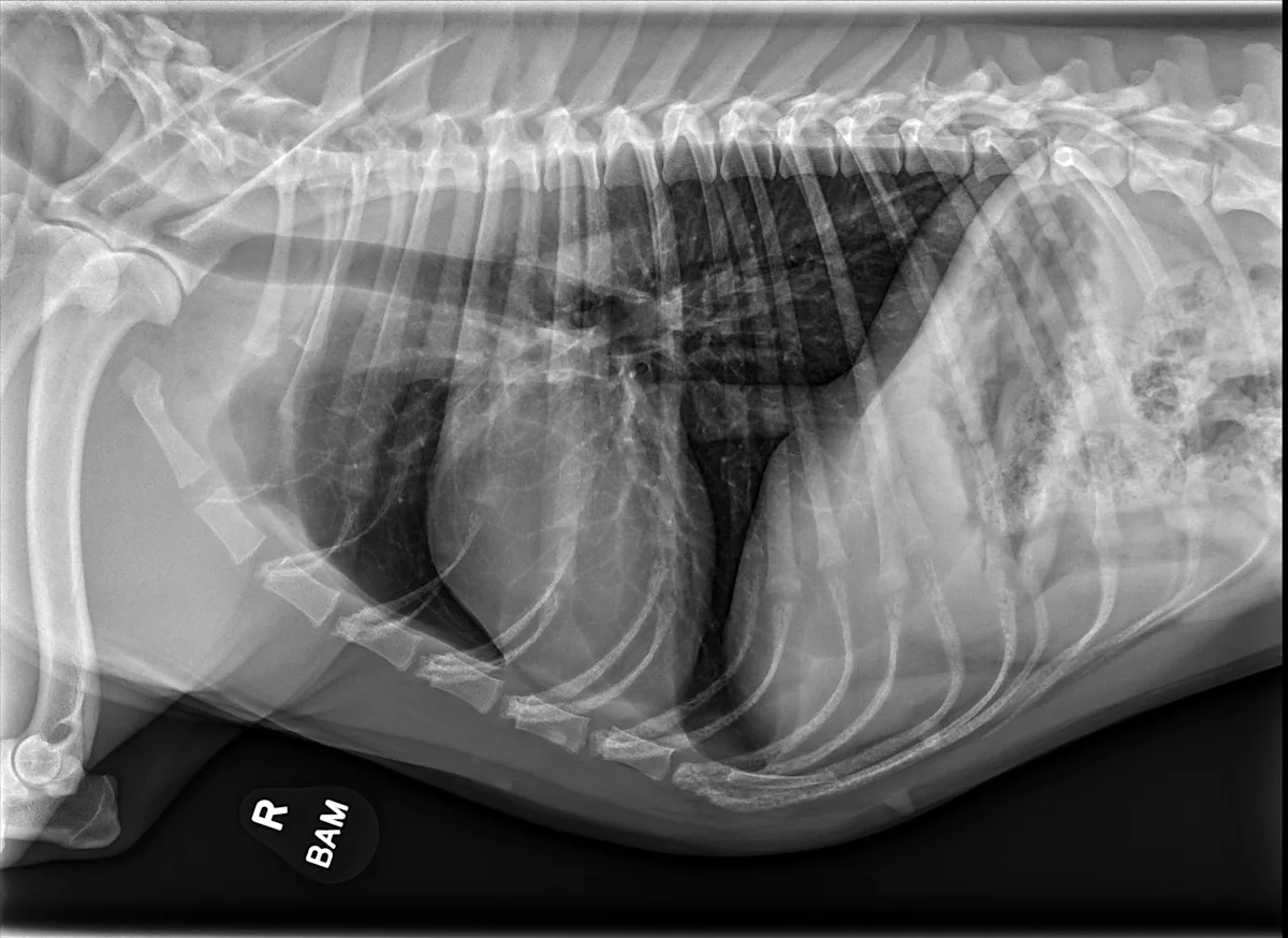
Not all cardiac changes are easy to spot on radiographs. This practical guide highlights key signs, explains limitations, and illustrates real-world cases of MMVD, DCM, PDA, and more.