Cardiac Library

Evaluation of the cardiovascular system involves more than simply listening through a stethoscope. Thorough evaluation also requires visual assessment of potential clinical signs, palpation to assess pulses and thrills, and clinical reasoning when evaluating patient history and signalment. This resource can aid clinicians seeking to elevate their knowledge of cardiac assessment, diagnosis, and management.

Sponsored By Sponsor Logo
A stylized illustration of a heart with a stethoscope positioned on it, symbolizing a cardiac examination. The heart is depicted in a light pink color, while the stethoscope is shown in blue, emphasizing the clinical focus on cardiovascular health.
A stylized illustration of a heart with a sound wave emanating from it, representing cardiac sounds. The design emphasizes the heart's anatomy and includes the text "Cardiac Sounds" in bold, red lettering below the heart.
A stylized illustration of a heart with an overlaid ECG waveform, highlighting the connection between cardiac anatomy and electrical activity. The design emphasizes the importance of ECG interpretation in veterinary medicine.
A stylized graphic of a heart wearing black headphones, symbolizing the connection between veterinary medicine and audio learning. The word "Podcasts" is prominently displayed below the heart, emphasizing the educational focus of the content.
A stylized illustration of a heart with question marks, suggesting a quiz or self-assessment related to cardiac health. The design features a gradient of pink hues and includes the text "Test Yourself" prominently below the heart.
A stylized illustration of a heart with a syringe and a medication bottle positioned inside it, symbolizing the concept of therapeutics in veterinary medicine. The background is a simple white, emphasizing the heart and its associated medical elements.
A dog resting.

Vertebral left atrial size (VLAS) is an objective radiographic measurement that can help determine whether left atrial enlargement is present with suspected or diagnosed myxomatous mitral valve disease. Review how to calculate VLAS in dogs.
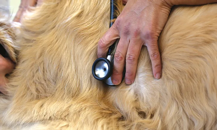
A golden retriever with a hand holding a stethoscope against its chest.

There are 12 components of a basic cardiology examination. Can you name them all?
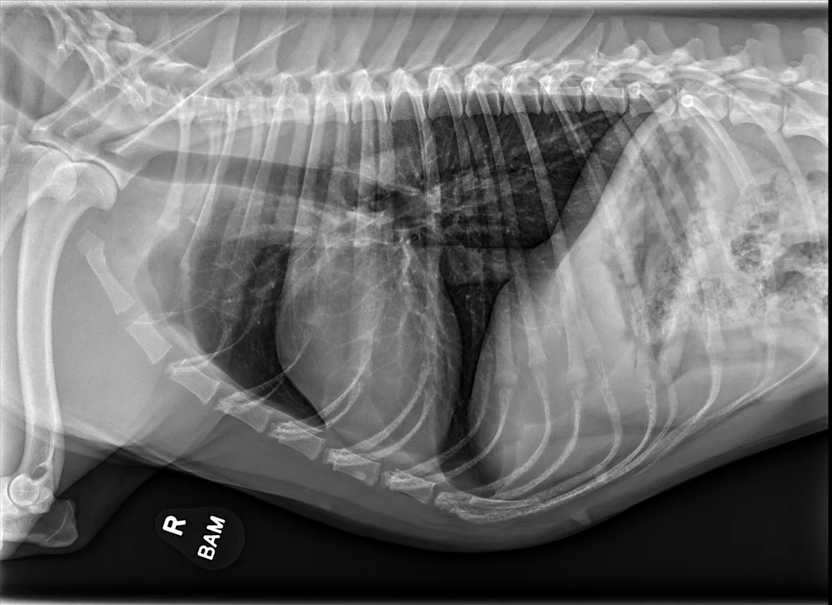
Radiograph in a dog.

Not all cardiac changes are easy to spot on radiographs. This practical guide highlights key signs, explains limitations, and illustrates real-world cases of MMVD, DCM, PDA, and more.