Drugs & Therapeutics
Learn about the latest veterinary medications and treatment options, including dosages, side effects, and interactions, from experts in the field.
Sponsored By 
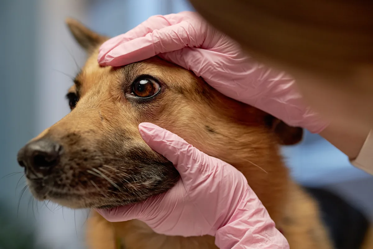
Using a natural language processing model, researchers analyzed a medical database to better understand the link between potentiated sulfonamide and KCS, including breed and age predisposition and the impact of dose and duration of treatment.
Topics In Drugs & Therapeutics
Featured in Drugs & Therapeutics


Poll
Quiz
New in Drugs & Therapeutics
Get more clinical guidance with Standards of Care™
From the team that brings you Clinician’s Brief, extend your knowledge with expert-written, peer-reviewed diagnostic and treatment guidance as well as pet owner education and all the reliable drug information you love in Plumb’s.