Lick Granulomas
Amelia G. White, DVM, MS, DACVD, Auburn University
Faith H. Scott, DVM, Auburn University
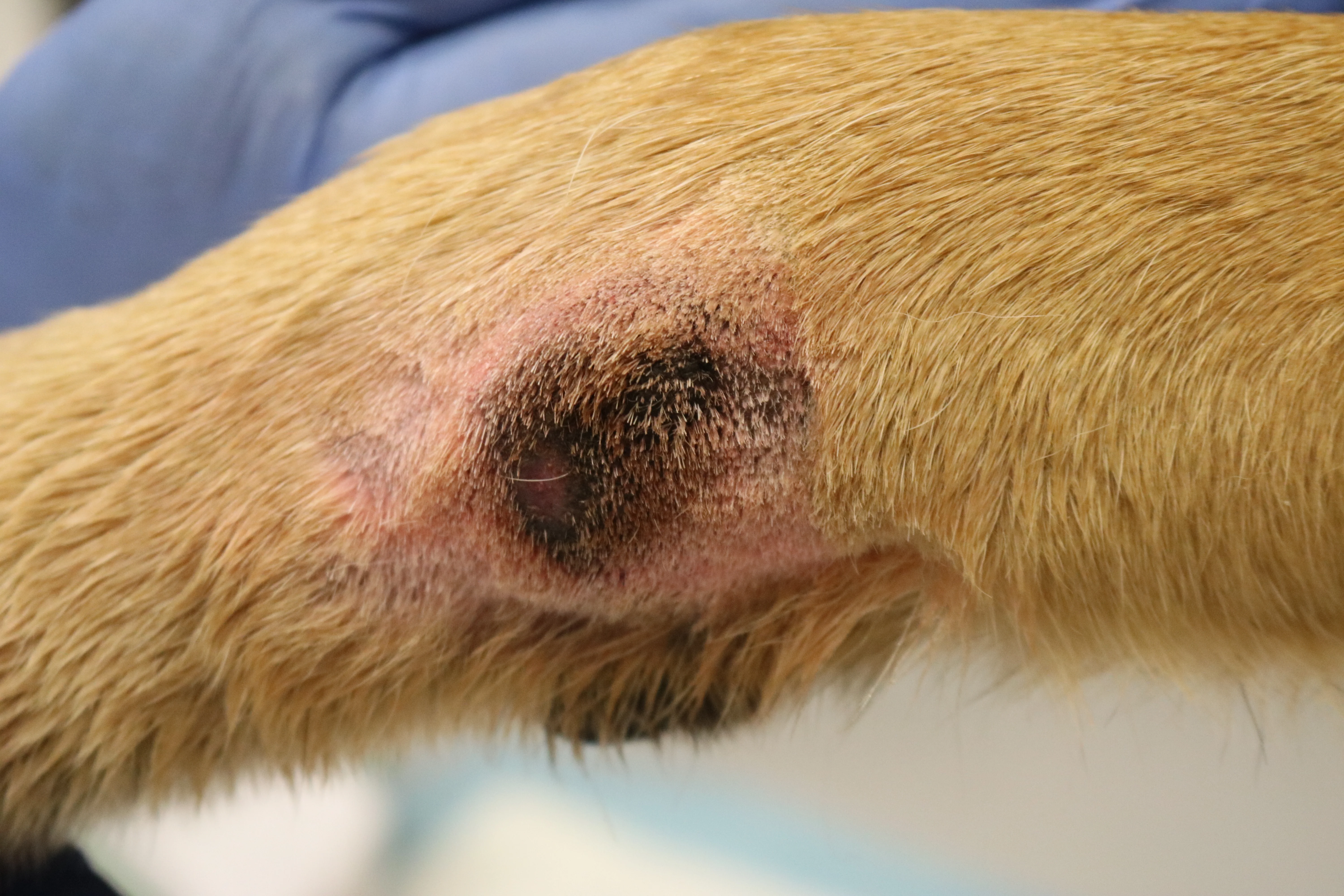
Lady, a 6-year-old spayed American Staffordshire terrier, was presented with a pruritic cutaneous mass on the right dorsal metatarsus of 2 years’ duration that had grown noticeably during the prior 6 weeks.
Physical Examination
On presentation, Lady was bright, alert, and responsive with normal vital signs. Physical examination was unremarkable; however, the right popliteal lymph node was mildly enlarged. A 5 × 3.5–cm well-demarcated, alopecic, raised, erythematous, ulcerative dermal mass withsanguinous, purulent discharge from drainingtracts was appreciated on the dorsal aspect of the right metatarsus. Additional dermatologic abnormalities included hypotrichosis in the tail gland region and mildly pruritic pododermatitis of the palmar/plantar aspects of all 4 paws characterized by moderate erythema and salivary staining.
Diagnosis
Differential diagnoses for a pruritic mass with draining tracts on the distal pes include acral lick granuloma/dermatitis (ALD), deep infection (eg, fungal, bacterial, Demodex spp), trauma, foreign body, furunculosis,and neoplasia.1,2 Unilateral lymphadenopathy raised concerns for a neoplastic or inflammatory process associated with the mass. Hypotrichosis of the tail gland area could be related to endocrinopathy (eg, hypothyroidism), demodicosis, pyoderma, or atopic dermatitis. The presence of pruritic pododermatitis supported a diagnosis of underlying atopic dermatitis.
Diagnostics
A complete patient history is needed to determine the underlying cause for accurate diagnosis. Important questions include age of onset, severity of pruritus, whether pruritus preceded lesion development, seasonality, indoor/outdoor lifestyle, ectoparasite prevention status for all animals in the home, whether any other animals or humans in the home have similar lesions,concurrent medications, known diseases, and travel anddietary history. In this case, physical examination findings and presence of the lesion for 2 years with significant seasonal worsening of pruritus supported atopic dermatitis as the primary cause.
Diagnosis should be confirmed and complicating factors (eg, orthopedic disease, foreign body, deep bacterial or fungal infection) assessed. Complete blood work and thyroid panel results were unremarkable. Fine-needle aspirate of the enlarged right popliteal lymph node was consistent with reactive lymphoid hyperplasia.
Radiography and skin diagnostics were performed with the patient under sedation via butorphanol (0.3 mg/kg IV) and dexmedetomidine (5 micrograms/kg IV). Radiographs of the metatarsus (Figure 1) revealed no osseous involvement. Direct impression skin cytology, skin scrapings, and tissue biopsy samples for histopathologic evaluation and tissue cultures (ie, aerobic, fungal) were collected. Direct impression smear and fine-needle aspiration results revealed RBCs, neutrophils, eosinophils, and lymphocytes without bacteria. No mites were observed on deep skin scraping. Biopsy sites were cleaned with chlorhexidine solution and infused with 2% lidocaine prior to sampling. Four 6-mm punch biopsies were collected for histopathology, aerobic culture, and fungal culture. A reversal agent (atipamezole, 0.5 mg/kg IM) was administered.3
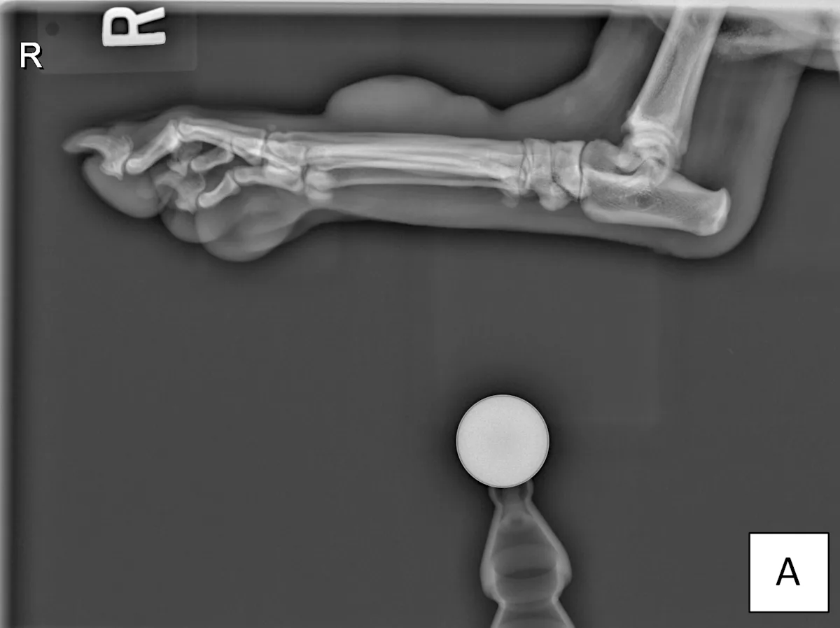
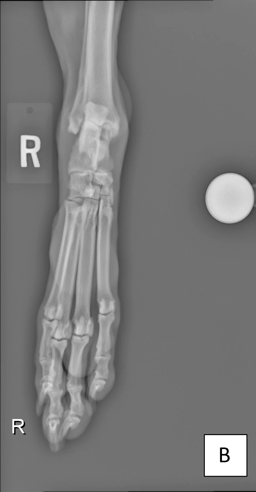
FIGURE 1 Lateral (A) and dorsoplantar (B) views of the right metatarsus. A smoothly marginated fusiform soft tissue cutaneous mass can be seen at the dorsal aspect of the right metatarsal region. No changes were identified in the underlying osseous structures, and the remainder of the study was unremarkable.
Fungal tissue culture was negative, and aerobic culture revealed light growth of Staphylococcus pseudintermedius with broad antibiotic susceptibility. Histopathology displayed marked epidermal parakeratotic and orthokeratotic compact hyperkeratosis and acanthosis, lymphoplasmacytic superficial dermatitis, marked dermal fibrosis, and dilated apocrine glands. Periodic acid–Schiff and Grocott’s methenamine silver staining can be used to visualize bacteria and fungi, respectively, on histopathology; however, these stains are not as sensitive as culture.
Diagnosis: Chronic Acral Lick Dermatitis Secondary to Atopic Dermatitis
Treatment & Long-Term Management
Pillars of treatment for ALD include breaking the itch–lick cycle with topical antiseptics and anti-inflammatory medication, targeted antimicrobial therapy to address deep pyoderma, deterring access to the site to prevent ongoing trauma from licking, and behavioral modification medications if a psychogenic component is likely.4 Surgical removal of ALD lesions has a high possibility of poor wound healing and lesion recurrence.5-8
Lady was prescribed a topical corticosteroid (fluocinolone acetonide in 60% dimethyl sulfoxide every 24 hours) to target the ALD lesion and lokivetmab (2 mg/kg SC once monthly) to target pruritus from the underlying atopic dermatitis. Fluorescent light therapy was administered for 2 back-to-back sessions on the same day every 7 days to decrease antimicrobial growth and inflammation and to accelerate wound healing (Figure 2).9 An Elizabethan collar was used to disrupt the itch–lick cycle.
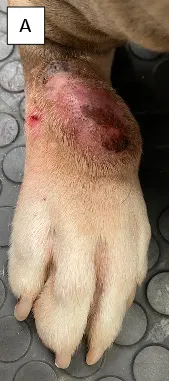
FIGURE 2 Healing progression of ALD on the right dorsal metatarsus of a dog treated with fluorescent light therapy on presentation (A) and on days 21 (B), 35 (C), 45 (D), and 60 (E)
Treatment at a Glance
The primary cause for licking (eg, pain, pruritus) should be identified and treated.
Thorough patient history, dermatologic examination with cytology, orthopedic examination, and radiography are standard.
The itch–lick cycle can be disrupted with administration of fast-acting antipruritic and anti-inflammatory medications (eg, corticosteroids, oclacitinib, ilunocitinib, atinvicitinib, lokivetmab).
Ongoing trauma can be prevented with lick barrier protection (eg, Elizabethan collars, leg wraps, bandages, clothing).
ALD is a form of deep pyoderma; tissue cultures can thus guide appropriate systemic antimicrobial selection when topical therapies are inadequate to resolve infection.14
Tissue samples for fungal culture and histopathology can rule out other factors that contribute to ALD lesions.
Topical therapy is an effective adjunctive treatment for infection and inflammation associated with ALD.
Early evidence suggests fluorescent light therapy may be an effective monotherapy or adjunctive treatment for control of infection and inflammation associated with ALD.9
Surgical resection of ALD is not a preferred treatment choice.
Outcome
Within 14 days, the size of the ALD lesion decreased by ≈72% from initial presentation and was nonpruritic after the third fluorescent light therapy treatment. At the 2-month recheck, moderate generalized erythema and pruritus were noted as associated with seasonal change. Atopic dermatitis was revisited, and oclacitinib (0.5 mg/kg PO every 24 hours) was prescribed to address allergic pruritus and inflammation. Weekly SC injections of allergen-specific immunotherapy were initiated and continued long term.
Discussion
ALD is a common but difficult to manage dermatopathy that accounts for nearly 3% of veterinary dermatologic conditions.10 A history of incessant focal licking, typically of the distal thoracic limb, can lead to a diagnosis of ALD.11 ALD was previously believed to be a primarily psychological condition, but lesions often begin with a stimulus that triggers the patient to lick constantly at the area, which may include a pruritic or painful signal.5,12 Management includes identifying and controlling the primary trigger for the itch/pain stimulus, treating complicating factors (eg, infection), preventing ongoing trauma from licking, and performing culture and susceptibility testing to guide treatment.1,5,12,13 A systematic approach can elucidate the contributing factors that require treatment, identify when behavioral modifying therapy is appropriate, decrease time to recovery, and prevent treatment failures.
Many ALD lesions are complicated by bacterial infection. A large discrepancy between superficial swab culture samples of an ALD lesion versus tissue cultures is typical, and poor response to empirical antibiotic treatment has been described.12 The International Society for Companion Animal Infectious Diseases antimicrobial guidelines categorize ALD as a deep pyoderma,14 and proper antimicrobial selection is crucial to reduce antimicrobial resistance.1 A 6-mm punch biopsy of a nonulcerated, sterile-prepared section of the lesion is standard, ensuring adequate depth for tissue culture and susceptibility.12 The antibiotic course recommendation is an initial duration minimum of 3 weeks with topical antimicrobials, followed by re-examination every 2 weeks until resolution; rarely, antibiotic courses are required for >6 weeks to eliminate secondary deep bacterial infection of ALD.14 Fluorescent light therapy uses the photobiomodulatory effects of light to reduce pyoderma healing time by 50% by decreasing inflammation, facilitating bacteria eradication, and hastening healing via stimulation of the natural healing process.9
Take Home Messages
The primary trigger for the itch/pain stimulus should be identified and controlled.
Ongoing trauma from licking should be prevented.
Complicating factors (eg, infection) should be treated.
Culture and susceptibility testing should be used to guide treatment.
A long-term plan to control the primary disease to prevent recurrence should be developed.