Fine-needle aspiration is a diagnostic procedure frequently used to evaluate cutaneous and subcutaneous masses, which are common findings during routine examination.1 Most skin masses appear to be benign; however, malignancy is possible. Cytology can provide a definitive diagnosis or narrow the category of differentials, helping determine whether the lesion is benign or malignant. Although cytology is usually consistent with the histopathologic diagnosis for skin masses, histopathology may be needed to assess malignancy and for definitive tumor typing due to overlapping cytologic features.1
Following are cytologic images of common skin tumors found in dogs and cats and their associated cytologic features.
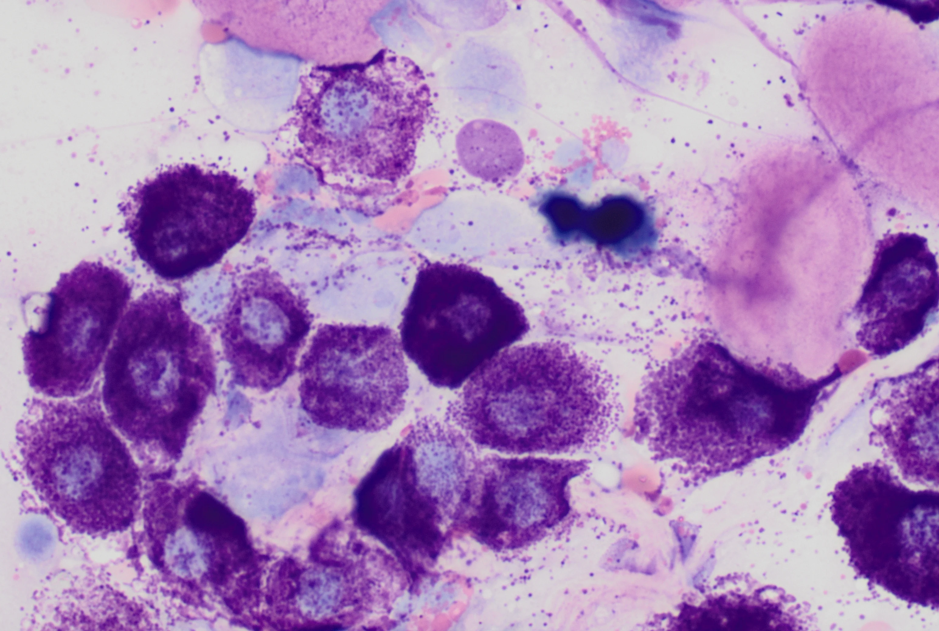
Basilar Epithelial Neoplasm

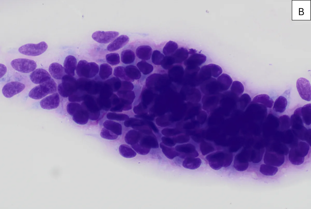
FIGURE 1 Cytology of a cutaneous mass on the lateral thorax of a 12-year-old spayed domestic shorthair cat (A); modified Wright-Giemsa stain; magnification, 200×. Cytology of a cutaneous mass from the cheek of a 14-year-old neutered male Pomeranian (B); modified Wright-Giemsa stain; magnification, 500×
Basilar epithelial neoplasia is an umbrella term for a group of skin tumors that structurally resemble basal epithelial cells and have a similar cytologic appearance.2 These are the most common skin tumors in cats and include trichoblastoma, pilomatricoma, and apocrine ductular adenoma.2-4 Basilar epithelial neoplasms are classically epithelioid in appearance and arranged in tight, dense, basophilic clusters with few individualized cells. Cells have a relatively uniform appearance, a cuboidal shape, and scant amounts of deeply basophilic cytoplasm that may be pigmented at times. Cells often appear lined up in rows within the clusters and are occasionally surrounded by pink fibrillar material. Some tumors have cystic portions that contain fluid and/or keratin.
Basilar epithelial neoplasms are usually benign but can become cystic, enlarged, and inflamed; can be locally invasive; and are rarely metastatic. Histopathology is needed for definitive tumor typing.
Follicular Cyst or Tumor
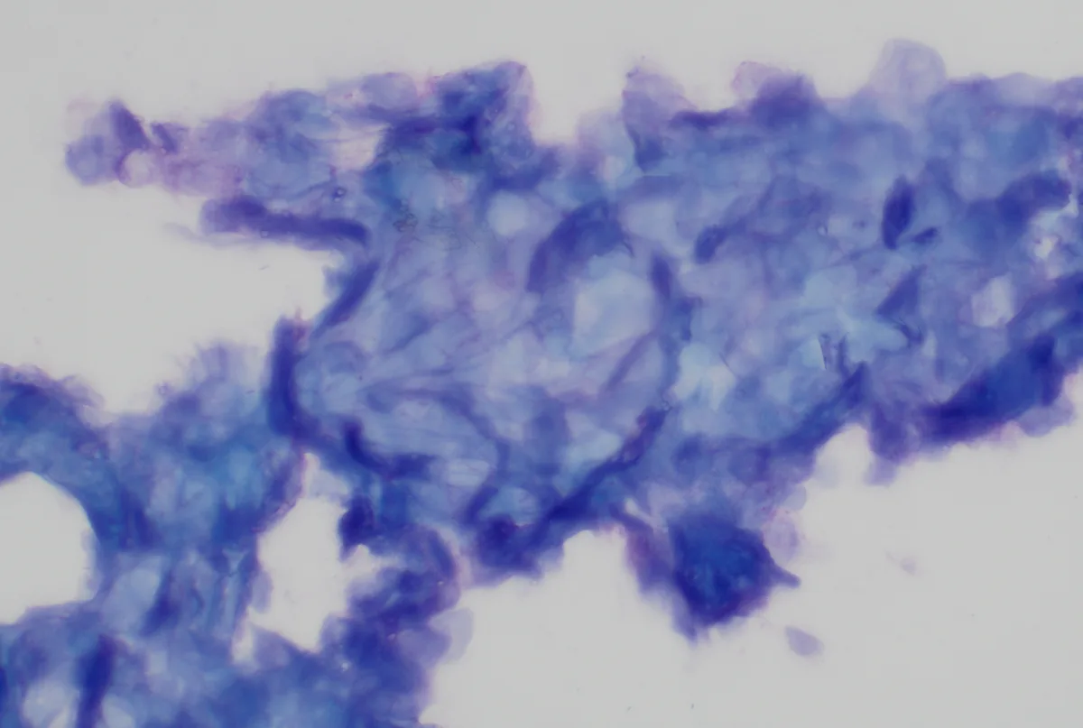
FIGURE 2 Cytology of a cutaneous mass from the cervical region of a 6-year-old intact male German Shepherd dog consistent with a diagnosis of follicular cyst or tumor; modified Wright-Giemsa stain; magnification, 100×
On cytologic examination, follicular cysts or tumors consist of mature, fully keratinized squamous epithelial cells and keratinaceous debris. Mature keratinized squamous cells are angular, flat cells that have lost their nuclei during maturation. The cytoplasm of the cells and cell fragments exhibit evidence of keratinization, which is characterized by a robin’s egg or sky blue color.
These lesions are usually benign but can become enlarged and inflamed. The presence of basaloid epithelial cells supports the diagnosis of a follicular tumor (eg, trichoblastoma)5; however, histopathology is needed to definitively distinguish cysts from tumors and for tumor typing.
Histiocytoma
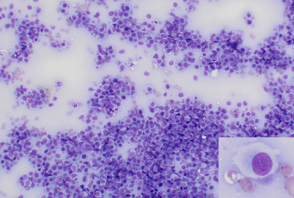
FIGURE 3 Cytology of a cutaneous mass on the paw of a 2-year-old neutered male crossbreed dog consistent with a diagnosis of histiocytoma; modified Wright-Giemsa stain; magnification, 100×. Cell with a pale, ruffled border (inset); magnification, 500×
Histiocytomas are the most common skin tumor in dogs <1 year of age but can be seen in dogs of all ages; some dogs may have multiple masses.6 On cytology, the tumor is composed of individualized round cells with variably well-defined cell borders and round, oval, or slightly indented nuclei. Cells have moderate amounts of pale staining cytoplasm; the outer edges of the cytoplasm demonstrate lighter stain uptake than the perinuclear area and may be ruffled in appearance. High numbers of small lymphocytes usually correlate with regression of the mass. Histiocytomas are typically benign, and spontaneous regression may occur in weeks to months.
Lipoma
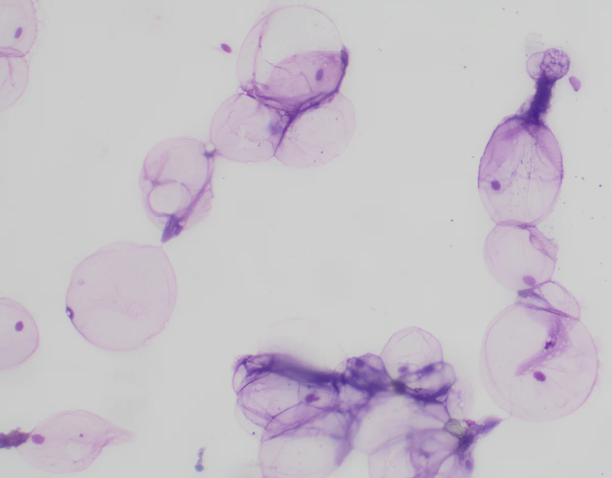
FIGURE 4 Cytology of a subcutaneous mass from the cervical region of a 9-year-old intact male Rottweiler consistent with a diagnosis of lipoma; modified Wright-Giemsa stain; magnification, 100×
Lipomas are benign tumors of adipose tissue that are common in dogs and can also be seen in cats.7 The adipocytes appear as aggregates of individualized, large, balloon-like cells with abundant pale-pink cytoplasm and a small, dark nucleus. These cells are easy to see on lower magnification because of their large size.
Adipose tissue has an oily appearance on the slide. Normal subcutaneous or perinodal adipose tissue may have a similar appearance. Alcohol fixatives used in Romanowsky-type stains can dissolve free lipid and may clear cells, making the sample appear acellular or nondiagnostic. Observing the slide grossly before staining can thus be helpful.
Canine Mast Cell Tumor

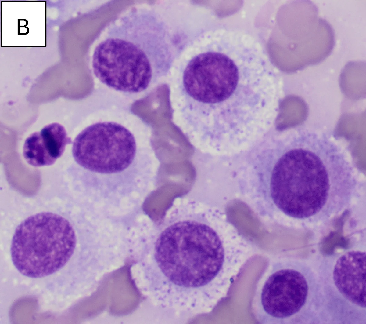
FIGURE 5 Cytology of a cutaneous mass from a 6-year-old spayed crossbreed dog consistent with a diagnosis of mast cell tumor (A); modified Wright-Giemsa stain; magnification, 500×. Mast cell tumor from a cutaneous hip mass from a middle-aged crossbreed dog (B); commercial Romanowsky stain; magnification, 500×. Although the granules do not stain well with this type of stain, a few granules can usually be appreciated at higher power.
Mast cell tumors are one of the most common skin tumors in dogs and cats.3,7 Sheets of round cells are observed on cytology. Well-differentiated cells are filled with fine pink/purple (metachromatic) granules that often obscure the nucleus; nuclear staining may be pale lavender. Eosinophilic inflammation is common in dogs and horses but uncommon in cats.8 Reactive mesenchymal cells (ie, fibroblasts) and pink collagen fibers may be seen. Fibroblasts can be prominent in some canine tumors and show cytologic atypia.
Romanowsky-type stains do not stain mast cell granules well, but these granules may be appreciated at higher magnification. Histopathology is needed to definitively grade mast cell tumors, as grade is correlated with prognosis in dogs.9,10 Some studies have used cytologic features to grade mast cell tumors; this practice may become more prevalent in the future.11
Feline Mast Cell Tumor
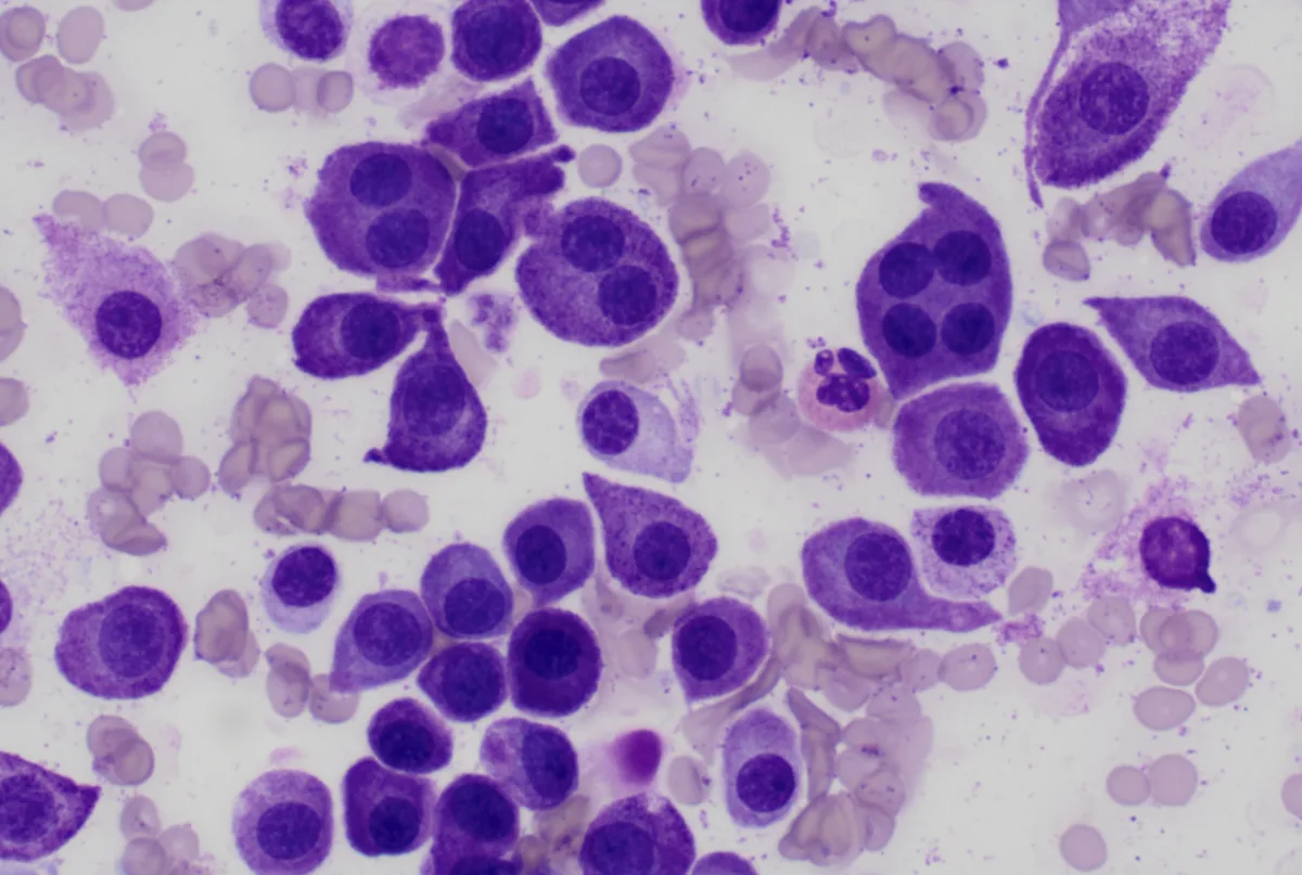
FIGURE 6 Cytology of a cutaneous mass on the pinna of a 10-year-old neutered male domestic shorthair cat consistent with a mast cell tumor; modified Wright-Giemsa stain; magnification, 500×
Cutaneous mast cell tumors in cats do not typically contain eosinophils, reactive fibroblasts, or collagen.8 Binucleated and multinucleated mast cells are more commonly observed in cats than in dogs. The histopathologic grading system used in dogs does not provide prognostic information in cats.8 A single cutaneous mast cell tumor in a cat is typically benign.8
Melanoma
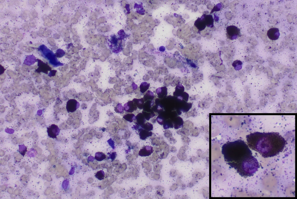
FIGURE 7 Cytology of a cutaneous mass in the lumbar region of a 7-year-old neutered male crossbreed dog consistent with a diagnosis of melanoma; modified Wright-Giemsa stain; magnification, 200×. Two oval-shaped, neoplastic melanocytes with round, eccentrically placed purple nuclei and a large amount of cytoplasm filled with numerous green–black granules (inset) magnification, ×500
Cutaneous melanomas are typically benign; however, histopathology is needed to assess malignancy. A cutaneous melanoma may be designated as a malignant melanoma on cytology if there is concurrent cytologic evidence of metastasis to a lymph node. Oral and subungual melanomas are more likely to be malignant in dogs. Cytologic appearance can be highly variable, as features of epithelial, mesenchymal, and round cells may be present. Pigmentation of the cells varies among different tumors and within individual tumors. Well-differentiated cells have abundant, fine melanin granules that appear green–black in color and are oval- to rice-shaped. Nuclear features of heavily pigmented cells may be obscured, causing cells to look like solid aggregates of pigment.
Plasmacytoma
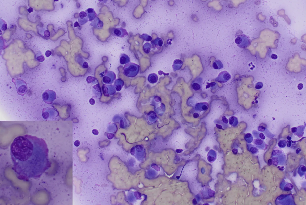
FIGURE 8 Cytology of a cutaneous mass on the paw of an 11-year-old neutered male Yorkshire terrier consistent with a diagnosis ofplasmacytoma; modified Wright-Giemsa stain; magnification, 200×. Flame cell with pink cytoplasmic border (inset); magnification, 500×
Plasmacytomas are not as common as other skin tumors (ie, <1% in dogs, rare in cats).7 Cells may be well-differentiated and easily recognized as plasma cells or may exhibit atypia. Cells are round with deeply basophilic cytoplasm that tends to stain darker at the margins and paler in the perinuclear zone, which is variably prominent. Nuclei are eccentrically placed and round with coarse chromatin. Binucleated cells are a common and consistent feature, and low numbers of multinucleated cells can also be seen. Plasma cells may have a bright pink cytoplasmic border (ie, flame cell).
Sebaceous Hyperplasia or Adenoma
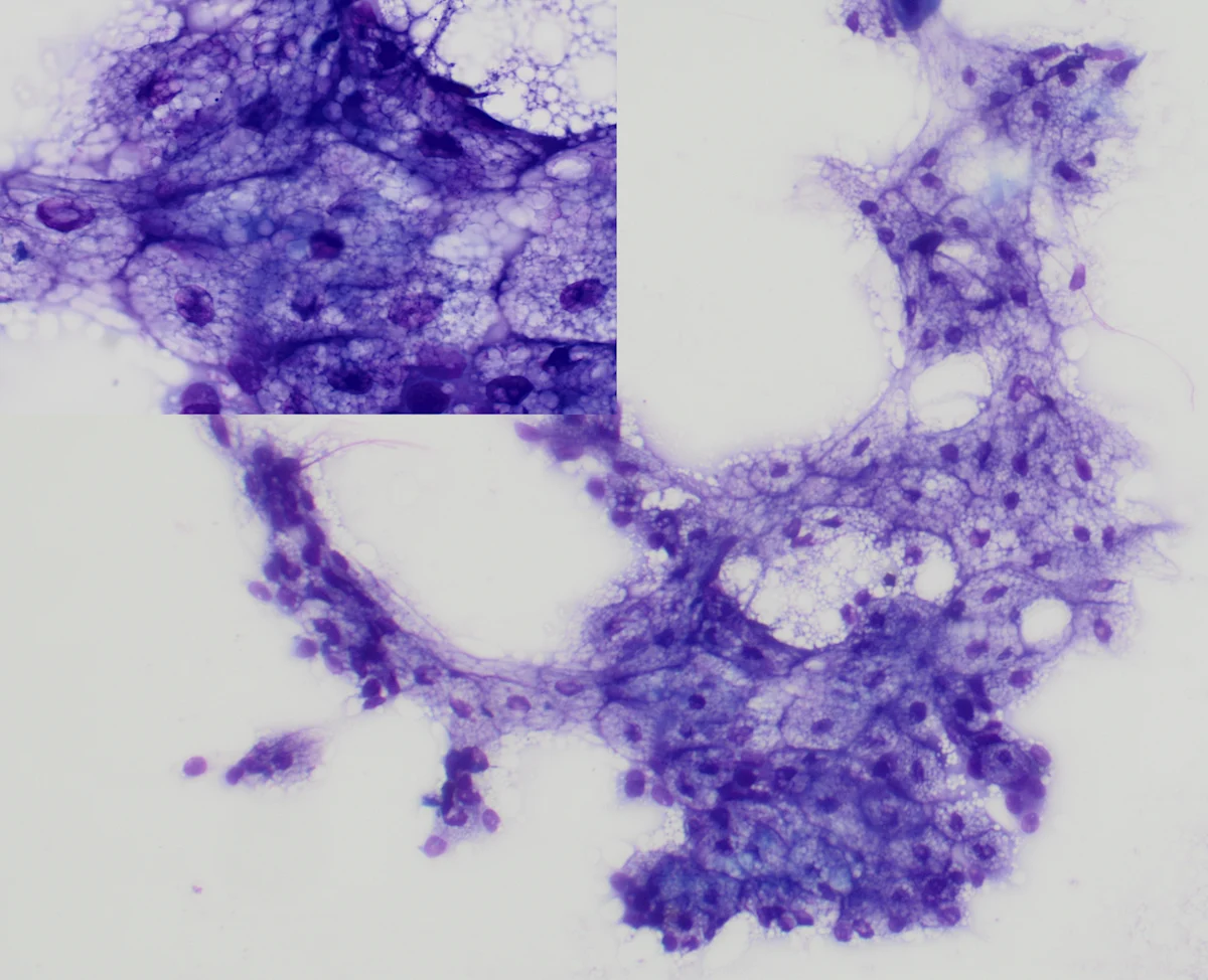
FIGURE 9 Cytology of a cutaneous mass on the hip of an 8-year-old neutered male crossbreed dog consistent with a diagnosis of sebaceous hyperplasia or adenoma; modified Wright-Giemsa stain; magnification, 100×. Moderate to abundant, highly vacuolated/foamy cytoplasm and small central nuclei (inset); magnification, 500×
Sebaceous hyperplasia or adenoma is a common lesion in older dogs and can be seen in cats. This lesion is composed of uniform, tightly cohesive clusters of well-differentiated sebaceous epithelial cells. Cells have moderate to abundant, highly vacuolated/foamy cytoplasm and small central nuclei. Small numbers of basal-appearing cells (ie, reserve cells) along the outer edge of the clusters may be apparent; these cells differentiate into mature sebaceous cells. The background of the slide is often vacuolated from sebaceous material released by ruptured cells. Histopathology is needed to differentiate sebaceous hyperplasia from sebaceous adenoma; however, both are benign.
Squamous Cell Carcinoma
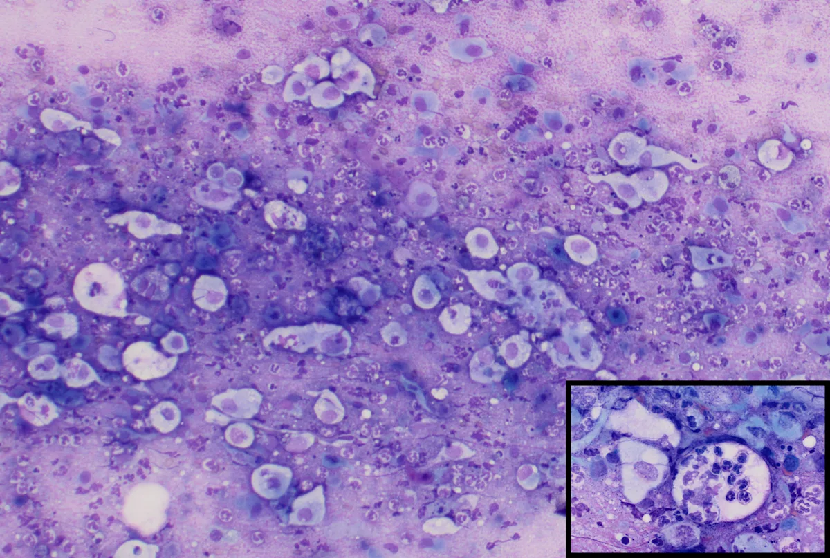
FIGURE 10 Cytology of a cutaneous mass on the head of a 9-year-old spayed crossbreed dog consistent with a diagnosis of squamous cell carcinoma; modified Wright-Giemsa stain;magnification, 100×. Neutrophils migrating through the neoplastic squamous cell (inset) magnification, 500×
Squamous cell carcinoma is one of the most common skin tumors in cats. Cytologic features include asynchronous nuclear to cytoplasmic maturation (mature cytoplasm appears as an angular shape with keratinized cytoplasm) with a large, round, immature nucleus.2 Other features include emperipolesis (ie, neutrophils migrating through the neoplastic squamous cell, tadpole‐shaped squamous cells, and small perinuclear vacuoles. Squamous cell carcinomas are frequently inflamed, and neutrophils exfoliate alongside the neoplastic cells, which may be present in clusters or individually.2 In nonneoplastic inflammatory lesions, squamous cells can become dysplastic and display features of malignancy.
Squamous Papilloma
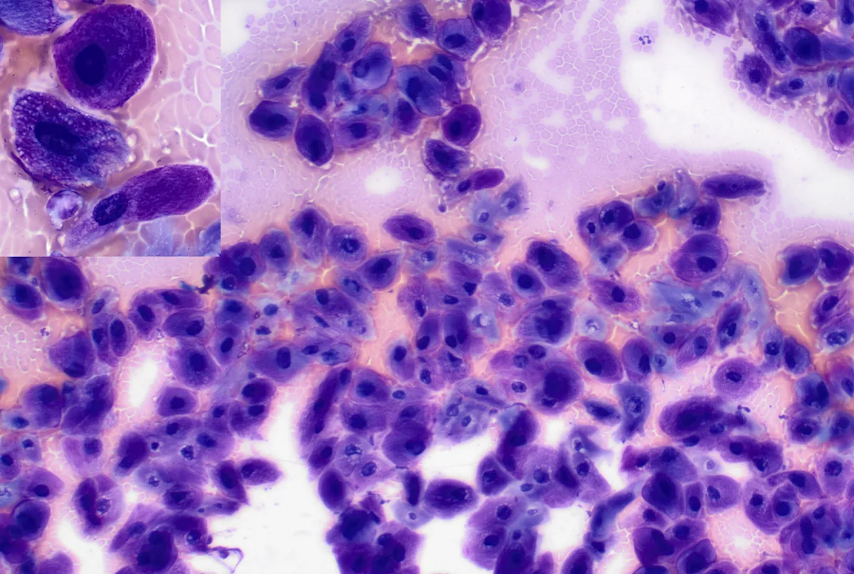
FIGURE 11 Cytology of a cutaneous mass on the paw of a 1-year-old neutered male Labrador retriever/poodle crossbreed consistent with a diagnosis of squamous papilloma; modified Wright-Giemsa stain; magnification, 100×. Characteristic dark-pink cytoplasm (inset) magnification, 500×
Papillomas are one of the most commonly diagnosed skin masses in young dogs.6 These lesions may or may not be associated with viral infections. The cells are polygonal to angular to fusiform in shape with light basophilic to dark-pink, foamy cytoplasm and a round eccentric nucleus with clumped chromatin.2 Squamous cells with dark-pink cytoplasm are referred to as koilocyte-like cells and are typically associated with viral infections. These lesions can regress spontaneously.