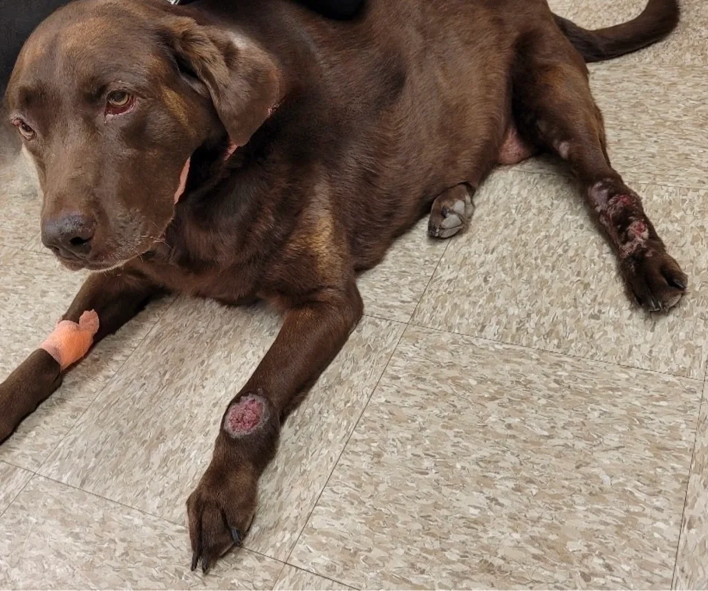
FIGURE 1 Acral lick dermatitis lesions on the left thoracic and pelvic limbs of a Labrador retriever
Acral lick dermatitis, a slow progressive condition induced by chronic licking, is more commonly diagnosed in large-breed dogs and affects the distal extremities of one or more limbs. This condition is clinically characterized as an erythematous, crusted lesion that progresses to alopecia, thickening of the skin, erosions, and ulcers. Exposure of deep layers of the epidermis, dermis, and sensory nerve fibers predisposes these dogs to deep pyoderma, ruptured hair follicles, and glands and induces further pruritus, perpetuating the itch–lick cycle.1 Dogs with acral lick dermatitis may develop a secondary compulsive disorder that can perpetuate the disease process.

FIGURE 2 Acral lick dermatitis seen as alopecia, erythema, and skin thickening on the right thoracic limb of a large crossbreed dog
Differential Diagnosis
Allergic dermatitis is the leading causative factor of acral lick dermatitis in most cases. Thorough patient history and dermatologic examination are essential for evaluation of potential underlying allergic disease. Pruritus on other areas of the body and recurrent skin and ear infections may also be present. Dogs with acral lick dermatitis should be considered to have allergic skin disease until proven otherwise. Lesions may not improve or can recur if underlying allergic disease is not properly addressed.
If allergic disease is ruled out, additional differentials can be considered, including pain associated with trauma, osteoarthritis, fracture or surgical sites, peripheral neuropathies, foreign body, primary psychogenic disorder, neoplasia, deep bacterial or fungal infections, and demodicosis.
Investigation
Thorough patient history is necessary to help identify the underlying cause of licking behavior and to inform workup and treatment. Different causes of allergies (eg, flea allergy dermatitis, food- or environment-induced atopic dermatitis) can be the primary cause of acral lick dermatitis. Investigation should include evaluation for signs of allergies (eg, pruritus, focal or generalized erythema, recurrent ear and skin infections). Depending on patient history and clinical findings, evaluation should include adding or changing flea/tick preventives, performing a strict dietary trial, and/or administering medical treatment for environmental allergies.
Diagnostics
Cytologic examination of the lesion is key in identification of secondary pyoderma. The lesion should be squeezed during collection of cytologic samples to express fluid from the deeper tissue to better ensure a diagnostic sample. Cytology can also help identify other conditions (eg, deep fungal infection, atypical bacterial infections, neoplasia) that can appear clinically similar.
Skin biopsy with histopathology can differentiate between a neoplastic process, underlying fungal disease, and atypical bacterial infection or support diagnosis of acral lick dermatitis. Further diagnostic testing is indicated if there are no other signs of allergic disease. Additional testing may include skin scrapings to rule out demodicosis and/or orthopedic examination to evaluate for joint disease. Diagnostic imaging is warranted if the patient has no signs of pruritus or allergies on other areas of the body and patient history suggests trauma or pain as the underlying cause.
Treatment
Acral Lick Dermatitis
Systemic glucocorticoids (eg, prednisone, starting dose of 1 mg/kg PO once every 24 hours) can effectively manage inflammation and pruritus associated with foreign body reaction to free keratin (furunculosis and contents of ruptured glands) and help reduce proliferative/fibrotic tissue.
Lesions may heal with scarring depending on chronicity and severity.
Long-term maintenance therapy for the underlying allergic disease should be discussed with the pet owner.
Carbon dioxide laser ablation should not be used as initial therapy without addressing the primary disease and secondary infection, as lesions will recur. This technique may be considered if lesions persist after potential underlying disease has been addressed and infection has resolved.1
Low-level laser therapy has not shown significant clinical efficacy as adjunctive therapy for acral lick dermatitis.
Secondary Pyoderma
Deep pyoderma is a bacterial infection that occurs in the dermis and sometimes extends into the subcutaneous tissue. Skin lesions indicative of deep pyoderma include erosions, ulcers, hemorrhagic crusts, purple papules, and nodules.2 Ninety-four percent of dogs with acral lick dermatitis have secondary deep pyoderma.3
Systemic antibacterial therapy is always indicated for deep pyoderma. The choice of antibiotic should ideally be based on bacterial culture and antimicrobial susceptibility testing. If bacterial culture cannot be performed and resistant infection is not suspected, first-choice antibiotics (amoxicillin/clavulanate, 12.5 mg/kg PO every 12 hours; cephalexin, 25 mg/kg PO every 12 hours; clindamycin, 11 mg/kg PO every 12 hours) can be administered empirically.2
Adjunctive topical antimicrobial therapy is recommended if a patient is considered pain free. Mupirocin 2% ointment/cream can be an option. Dimethyl sulfoxide (DMSO) gel can be administered immediately prior to mupirocin application to increase antibiotic penetration into the lesion. Owners should be instructed to wear gloves when administering topical medications and to immediately wash their hands.
An initial, 3-week course of systemic antibiotics should be prescribed and the patient rechecked with cytologic re-evaluation before the end of the 3-week course. If the bacterial infection is improving but not resolved on cytology, treatment should be continued with re-evaluation every 2 weeks.2
Additional Considerations
Physical restraint (eg, an Elizabethan collar) can be used temporarily. After the first few days of treatment, physical restraint should be removed for short periods with supervision. The restraint should only be removed without supervision after the patient has stopped licking.
Environmental factors (eg, excessive confinement, lack of exercise) that can be improved should be addressed. Selective serotonin reuptake inhibitors can be considered in conjunction with allergy treatment if behavioral conditions are potential or known components.