Canine Interdigital Follicular Cysts
Darren Berger, DVM, DACVD, Iowa State University
Ask the Expert: What do I need to know about diagnosing and treating interdigital follicular cysts in dogs?
Pododermatitis is a generic term that describes inflammatory conditions that afflict the paws of dogs and is a common reason for presentation to the clinic. Several primary conditions can cause this condition, which may result in chronic recurrent pododermatitis manifesting with interdigital swelling and draining tracts (see Medical Conditions Associated With Interdigital Pododermatitis in Dogs).1,2 Clinical presentation mirrors recurrent otitis, with primary causes, predisposing variables, and perpetuating factors complicated by secondary infections. Early appropriate intervention and prevention can avoid significant chronic changes and fibrosis that can result in complications. Although pododermatitis in these conditions represents a clinical manifestation of a more extensive disease, lesions are confined strictly to the paws in some patients, which may be a result of interdigital follicular cysts. This condition is often mistaken for a foreign body or nonresolving pyoderma and can be a source of frustration for pet owners and clinicians.
Medical Conditions Associated With Interdigital Pododermatitis in Dogs
Atopic dermatitis or cutaneous adverse food reaction
Contact dermatitis
Deep or systemic mycoses (eg, phaeohyphomycosis, blastomycosis)
Dermatophytosis
Endocrinopathy
Foreign body
Hepatocutaneous syndrome (ie, superficial necrolytic dermatitis)
Hookworm dermatitis
Interdigital follicular cysts
Malassezia spp dermatitis
Neoplasia (eg, squamous cell carcinoma)
Pyoderma
Clinical Signs
Interdigital follicular cysts often appear as noticeable dorsal lesions accompanied by licking, chewing, or lameness but can also be perceived by owners as a cosmetic flaw. Lesions are usually first noted in young adult dogs (1-3 years of age) and appear to be more common in English bulldogs, other bull terrier breeds (eg, boxers, pit bull terriers), Labrador retrievers, golden retrievers, and dogs with conformation deformities.3-6
On physical examination, dorsal interdigital erythema, swelling, nodules, hemorrhagic bullae, and/or draining tracts are visible (Figure 1). The dorsal interdigital lesion directly corresponds to an area of alopecia, erythema, edema, and comedone formation on the ventral palmar or plantar surface skin between the digital and metacarpal or metatarsal pads. Interdigital follicular cysts most often occur on the front paws between the fourth and fifth interdigital space5,6; however, lesions may occur anywhere, affect >1 interdigital space, or occur symmetrically depending on the underlying conformational disturbance.
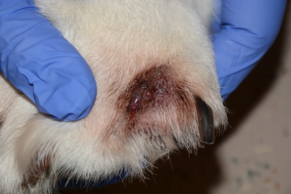
FIGURE 1 Nodule with a draining tract in the dorsal interdigital space between the fourth and fifth digits commonly encountered in patients with interdigital follicular cysts
Review the hallmark clinical signs, likely pathogenesis, and current treatment strategies for Feline Plasma Cell Pododermatitis.
Interdigital Follicular Cysts: What Are They & Why Do They Occur?
Interdigital follicular cysts form secondary to abnormal friction or trauma to the ventral interdigital webbing associated with congenital (varus or valgus deformity) or acquired anatomic deformities, osteoarthritis affecting weight-bearing mechanics, or uncontrolled dermatologic disease (eg, allergic dermatitis) resulting in conjoined paw pads or pseudopads.1,5,6 The abnormal friction creates thickened, edematous, or calluslike changes to the interdigital skin, resulting in plugging of follicular openings and comedone formation. Keratin production within the plugged follicular lumen persists and results in follicular dilation and cyst formation.5
Because dogs have compound hair follicles (ie, multiple hair shafts arising from 1 opening), a single plugged follicular opening can result in multiple cysts. Follicular cysts may thus be present in multiple layers of the ventral interdigital skin, and each is prone to rupture. Follicular rupture can result in release of keratin, hair, and bacteria into the dermal space, inciting a pyogranulomatous foreign body (eg, broken hair shaft) reaction along with secondary deep pyoderma. Concurrent deep pyoderma and foreign body reaction leads to incomplete resolution of lesions with antimicrobial therapy alone; inflammation reoccurs quickly once anti-inflammatory therapy is discontinued. Repeated rupture of the follicular cysts typically results in fistulous tract formation, which drains into the dorsal interdigital space. A dorsal clinical lesion commonly results in misinterpretation of the lesions originating at this site.5
Diagnosis
Interdigital follicular cysts are usually diagnosed based on clinical presentation and physical examination findings. Dermatologic samples should be acquired via cytology, skin scraping, and trichogram to eliminate other primary or confounding causes. To support diagnosis, pressure can be applied to the corresponding dorsal surface during examination of the palmar or plantar surface; extrusion of keratin from the plugged follicular openings usually results (Figure 2). Caution is needed for nonsedated patients, as a pain response may be elicited. Biopsy from the palmar or plantar surfaces (where interdigital follicular cysts originate) with histopathology can help confirm the diagnosis in difficult or confounding cases.
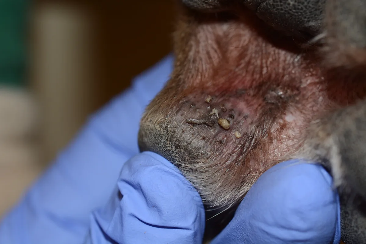
FIGURE 2 Palmar lesion corresponding to a dorsal interdigital swelling with extrusion of white-to-gray–colored keratin from plugged follicular openings following pressure applied to the dorsal surface
Get Your Skin Deep Dermatology Guide
Do you feel comfortable performing a trichogram to look for Demodex spp? What about skin scraping for Sarcoptes scabiei? You have the tools to perform the tests you need already in the clinic. Our free e-book, A Clinician's Guide to Dermatology, walks you through 5 common procedures you can do in-house.
Treatment
Surgical ablation is the preferred treatment for lesions; traditional surgery can be performed if medical management fails.4-9 Partial or total fusion podoplasty has been described for chronic, nonresolving interdigital dermatitis but should be considered a salvage procedure7,8; postsurgical complications include dehiscence, lameness, and, specifically with partial podoplasty, alteration to conformation and development of new lesions at a different interdigital site.
Surgical Ablation
Surgical excision and/or ablation with a carbon-dioxide laser is the preferred treatment for interdigital follicular cysts, is well-documented, and typically results in a first-time success rate of roughly 80%.4-6 Appropriate training and experience in performing this procedure are important, as clinical success depends on skill level; referral should be considered when needed.
Following surgical ablation, the treated area should be allowed to heal via secondary intention (Figure 3). At the time of writing, there are no consensus guidelines for postoperative care. Anecdotally, the following basic principles are used:
Topical antimicrobials (eg, silver sulfadiazine cream, manuka honey, fusidic acid, mupirocin, chlorhexidine)
± systemic antimicrobials
Postoperative pain control (eg, NSAIDs, opioids)
Exercise restriction
Bandaging (wide practice variations)
General protection of the site until healed
Mean healing time is 34 days (range, 22-46 days).6 Recurrence and repeated treatment procedures are typically needed in up to 10% of patients to permanently address the condition.5,6
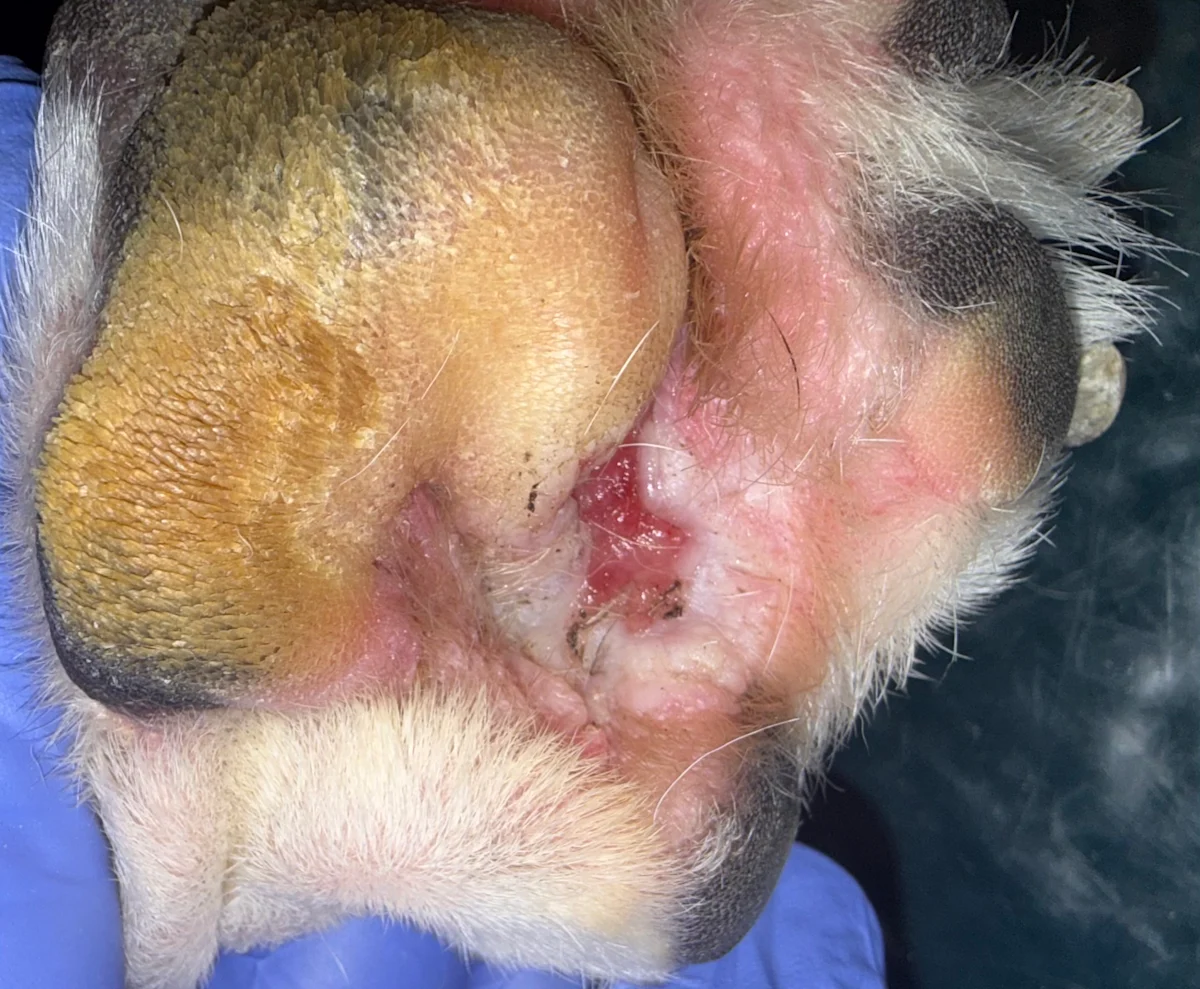
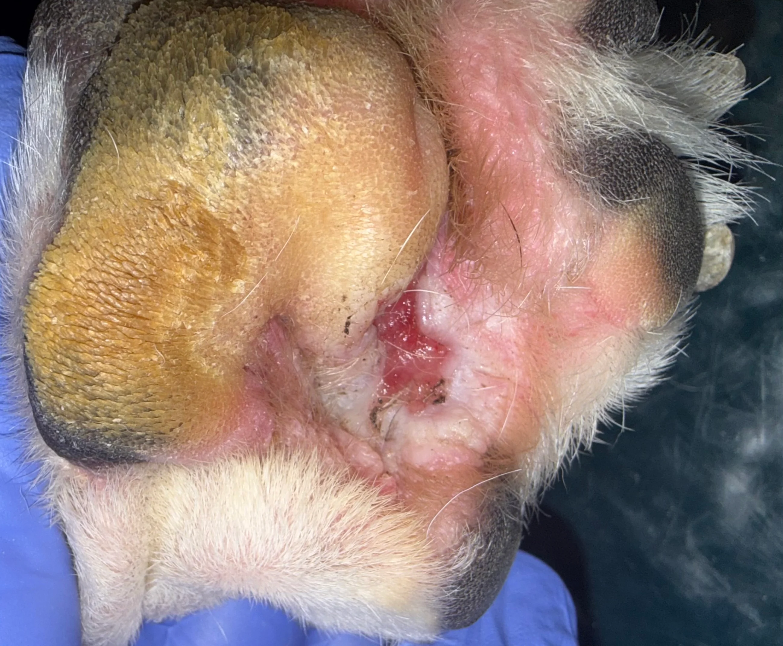
FIGURE 3 Second intention healing several weeks after surgical ablation with a carbon-dioxide laser. Image courtesy of J.B. Peiper, DVM, DACVD, Iowa State University
Medical Management
Medical management can be attempted in cases of early lesion development or when surgical intervention is not an option. In cases of interdigital follicular cysts, topical products (eg, gels, lotions, wipes) containing benzoyl peroxide or salicylic acid applied to the palmar or plantar aspect may be beneficial because of their follicular flushing and keratolytic properties. These products should be applied daily to every other day to break down and prevent comedone formation. Anti-inflammatory (eg, cyclosporin, Janus kinase inhibitors, glucocorticoids) and antimicrobial therapy may be beneficial in resolving secondary infections and inflammation associated with ruptured cysts. Fluorescence biomodulation may lead to quicker resolution of secondarily infected lesions when combined with systemic antibiotics and have delayed recurrence rates compared with antibiotic therapy alone.10
Medical treatment of infection and inflammation alone, without surgery, will not resolve this condition or prevent further episodes. Medical therapy has limited efficacy in most cases and is unlikely to be beneficial in chronic relapsing cases in which scarring has occurred.