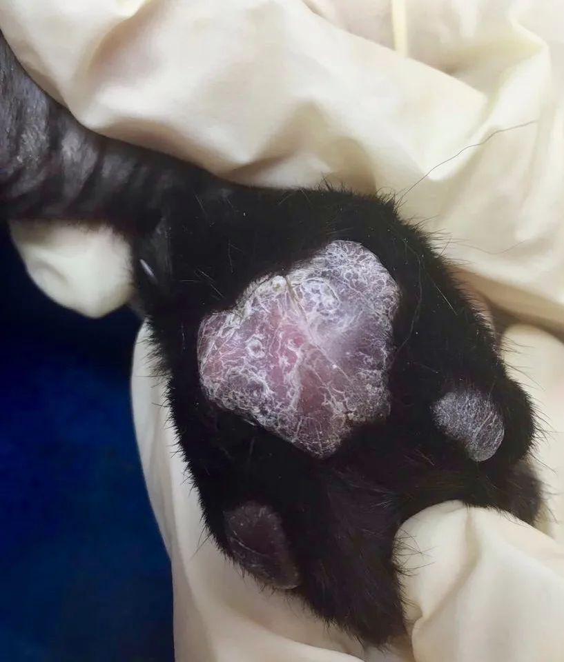
Appearance
Plasma cell pododermatitis is a rare dermatologic condition in cats characterized by soft swelling of the paw pads that may eventually ulcerate, causing pain and lameness.1-4 This condition has a characteristic clinical appearance of soft spongy swelling (commonly referred to as pillow foot), pink or sometimes violet purple color, white striations, ulceration, and loss of paw-pad architecture.1,3-6 Multiple paw pads are typically affected, with lesions predominantly on the central metacarpal and metatarsal pads; rarely, a single paw pad is affected.
Clinical Signs
Lesions are often painful. Some affected cats are presented with lameness; others may be subclinical.2,6 Additional possible clinical signs include poor body condition, hypersalivation, lymphadenopathy, pyrexia, anorexia, lethargy, and (rarely) plasma cell dermatitis with swelling of the nose or stomatitis.1,6 Immune-mediated glomerulonephritis and renal amyloidosis have also been reported in pododermatitis cases.1,7 Chronic lesions may be fibrotic. Hematologic findings include mature neutrophilia, leukocytosis with mild left shift, thrombocytopenia, monocytosis, and hypergammaglobulinemia. Anemia and lymphopenia have also been reported.1,2 Polyclonal gammopathy is present in the majority of cases and can persist after treatment.1,8
Differential Diagnosis
The cause and pathogenesis of plasma cell pododermatitis have not been completely elucidated; an immune system dysfunction seems most likely based on the positive response of many cats to immunomodulatory therapy, marked plasma cell tissue infiltrate, and hypergammaglobulinemia observed in most affected cats.1,2,4 Some patients, however, have seasonal relapses, suggesting environmental allergies as a possible cause.2 Infectious etiology, specifically FIV, has also been suggested.2,6 Studies have found 44% to 62% concurrent FIV positivity in cats with plasma cell pododermatitis6,7; however, whether FIV positivity has a role in pathogenicity of plasma cell pododermatitis is undetermined.1
Differentials may include eosinophilic granulomas/plaques, bacterial or fungal granulomas, foreign body reactions, nocardiosis, and leishmaniosis affecting the paw pads.1,2 A recent study that evaluated silica-based litter granulomas found that 10 out of 13 biopsies also contained evidence of plasma cell pododermatitis.9 Although the relationship between cat litter granulomas and plasma cell pododermatitis requires more investigation, the authors recommended permanently avoiding silica-based litter for cats that develop plasma cell pododermatitis.9
Tentative diagnosis is based on patient history, skin lesions, and a large number of plasma cells on fine-needle aspiration, cytologic examination, or skin biopsy. Cats with plasma cell pododermatitis may or may not be FIV and/or FeLV positive; a screening test is recommended for all patients diagnosed with plasma cell pododermatitis.1,2,6
Treatment
Historically, first-line treatment has included oral cyclosporine or oral doxycycline. Doxycycline and related antibiotics have immunomodulatory properties that can result in complete remission in one-third of cats and improvement in lesions in 80% of cats2,7; however, due to antimicrobial stewardship and risk for bacterial resistance, use of an antibiotic for nonantimicrobial purposes has raised controversy on whether these drugs are appropriate in specific cases.1 One study using immunohistochemistry and PCR failed to identify doxycycline-responsive pathogens.10
Oral cyclosporine (7 mg/kg every 24 hours until remission), a calcineurin inhibitor, is another immunomodulatory option for controlling the disease and may be preferred for antimicrobial stewardship.1
Depending on disease severity, systemic oral glucocorticoids (prednisolone, 2.2-4.4 mg/kg every 24 hours; triamcinolone acetonide, 0.4-0.6 mg/kg every 24 hours; or dexamethasone, 0.2 mg/kg every 24 hours) administered alone or in conjunction with other immunomodulatory treatments may be initiated in some patients.1,2
Once remission is achieved, medication can be gradually tapered then discontinued. Treatment should be resumed in cases of relapse. Surgical excision of affected paw pads is usually curative and reserved for cases that do not respond to medical therapy.1,2,4
Prognosis
Prognosis varies, as some patients may experience spontaneous resolution of clinical signs and others may require immunomodulating agents and lifelong therapy.1,2,4