In the Literature
Klim EB, Mestrinho LA, Gawor JP. Effect of a buried knot in the healing process of dental extraction sites: a prospective study in cats. J Feline Med Surg. 2025;27(3):1098612X251314701. doi:10.1177/1098612X251314701
The Research …
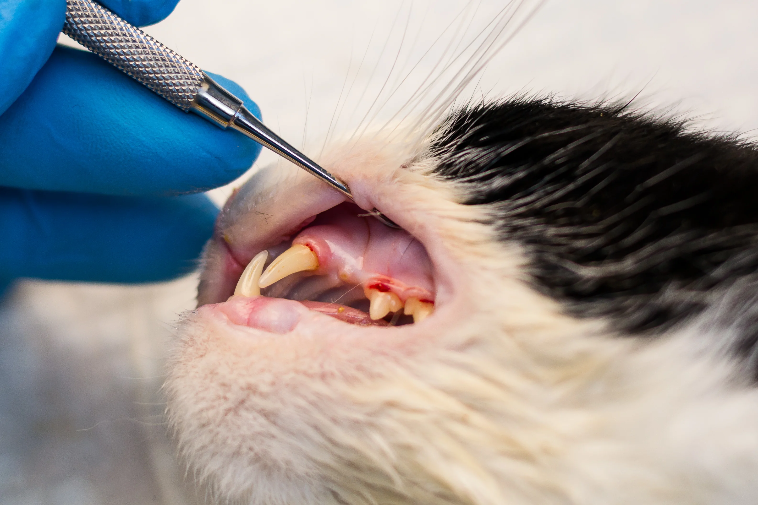
In general practice, periodontal disease has been reported to be the most diagnosed disease in cats.1 Periodontal disease and tooth resorption may result in loss of tooth structure.2 Extraction of an affected tooth is warranted when teeth incur significant attachment loss or loss of hard or soft tissue structures. The wounds caused by surgical extractions are commonly closed with an absorbable suture material and a simple interrupted, cruciate, or simple continuous pattern.3
This study evaluated the effectiveness of a buried-knot technique as an alternative to standard knot placement in a simple interrupted suture pattern. Forty predominantly domestic shorthair cats (mean age, 6.4 years) were presented for anesthesia and dental extractions for periodontal disease, tooth resorption, or gingivostomatitis. Each cat was identified as needing extractions performed in all 4 quadrants.
Surgical extractions were performed similarly with a mucoperiosteal flap, followed by postextraction radiography to verify removal of root remnants. A 5-0, absorbable, monofilament material with a tapered needle was used for closure. The gingiva of the maxillary and mandibular extraction sites on the ipsilateral side was closed using the buried-knot technique, and the contralateral maxillary and mandibular extraction sites were closed using standard simple interrupted sutures, allowing each cat to act as the test and control.
A surgeon’s knot combined with 4 simple knots was placed 3 mm apart and 2 mm from the wound edges at all extraction sites. Buried knots were placed by directing the needle from within the wound to the outside of the flap, followed by insertion of the needle outside of the wound from the opposite wound edge back into the wound, thus inverting the knot. All patients were administered the same analgesic and antibiotic protocol to reduce external factors that may affect healing.
Conscious evaluations were performed on all patients after 2, 4, and 6 weeks for swelling, bleeding, erythema, dehiscence, ulceration, discharge, pain, necrosis, flap instability, loose sutures, and debris accumulation on the suture material.
No significant difference was seen between the groups in relation to dehiscence, necrosis, flap instability, or loosening of sutures. The buried-knot group yielded less swelling, bleeding, erythema, ulceration, pain, and debris during the 2- and 4-week evaluations. At the 6-week assessment, no difference was observed between groups. Although 2 cats with chronic stomatitis showed dehiscence at the 2-week re-evaluation, the small sample size prevented demonstration of statistical relevance among groups of cats with periodontal disease, tooth resorption, or gingivostomatitis.
… The Takeaways
Key pearls to put into practice:
The buried-knot technique may be considered for dental extraction sites; simple interrupted sutures and buried knots to close dental extraction sites yield similar healing success.
Compared to simple interrupted sutures, buried knots may result in decreased erythema, swelling, and debris collection at the extraction site and fewer healing complications.
The buried-knot technique is not recommended in cats with chronic gingivostomatitis, as the persistent inflammation seen in this condition may reduce suture strength and result in dehiscence.
You are reading 2-Minute Takeaways, a research summary resource presented by Clinician’s Brief. Clinician’s Brief does not conduct primary research.