Periodontal Disease: Alternatives to Extraction
Kendall Taney, DVM, DAVDC, FAVD, Center for Veterinary Dentistry & Oral Surgery, Gaithersburg, Maryland
In the Literature
Alterman JB, Huff JF. Guided tissue regeneration in four teeth using a liquid polymer membrane. J Vet Dent. 2016;33(3):185-194.
The Research …
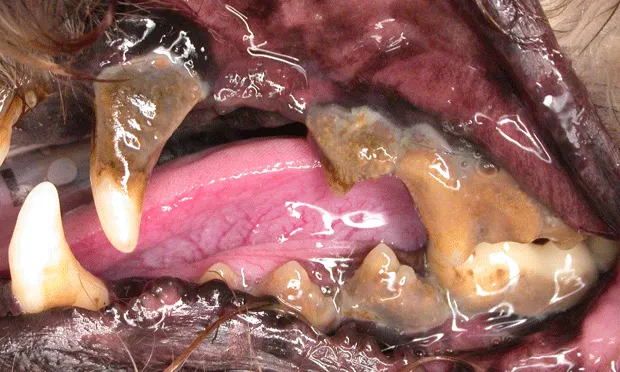
This case series discussed guided tissue regeneration (GTR) and its use in treating teeth affected by periodontitis and subsequent tissue loss. The goals of GTR are to reverse alveolar bone loss and re-establish attachment of cementum to alveolar bone via a healthy periodontal ligament.
Periodontal ligament cells must be the first tissue type to repopulate along the root surface for a pocket to successfully heal. Use of a barrier or membrane between the gingival tissues and the root surface facilitates repopulation of periodontal ligament, cementum, and bone cells. Without a membrane and/or proper debridement, epithelial cells may regenerate along the root surface and result in a long junctional epithelium and recurrence of periodontal disease and attachment loss. Alternatively, if bone cells are the first to repopulate the root surface, resorption or ankylosis can occur.
This paper described 4 teeth from 3 dogs with attachment loss ranging from 25% to more than 50%. The teeth were treated with open flap surgery to allow for debridement of the root surface and bony and periodontal pocket surfaces.

A Marquis periodontal probe with 3-, 6-, 9-, 12-, 15-, and 18-mm markings

Measurement of the gingival sulcus with a Marquis probe. Normal sulcar depth should be approximately 2 mm.
The authors used a commonly available polymer gel (doxycycline in a polymer-delivery system) to mimic the properties of an absorbable barrier membrane. The gel was placed between the flap and demineralized bone matrix graft material, which had been placed in the bony defect to encourage new bone growth. Based on dental radiography and probing six months postoperatively, all 4 teeth had attachment gain and new bone formation. Without histopathologic analysis, it was not possible to determine which cells had repopulated the root surface. However, an intact periodontal ligament was visible radiographically in each of the 4 teeth.
… The Takeaways
Key pearls to put into practice:
Each patient should be anesthetized for a thorough oral examination that includes probing of all teeth along multiple points. A dental probe with measured markings can identify problem areas (normal probing depth, ≈2 mm).
Basic GTR principles can be applied in general practice to improve attachment loss in smaller periodontal pockets (<4 mm). Closed root planing can be performed with a periodontal curette to debride all surfaces of the pocket. A perioceutic agent that delivers higher local concentrations of an antibiotic to the environment can be placed to facilitate healing and reduction of the pocket.
Extraction is not the only option. For strategically important teeth (ie, canines, carnassials) with deeper periodontal pockets (>4 mm), referral to a board-certified veterinary dentist should be considered if tooth preservation is desired. Open flap surgery and root planing followed by GTR can reverse attachment loss. For these procedures to be successful, owners must be dedicated to diligent home dental care.