Mandibular Extractions in Cats
Mark M. Smith, VMD, DACVS, DAVDC, AVDC and ACVS Founding Fellow of Oral & Maxillofacial Surgery, Center for Veterinary Dentistry & Oral Surgery, Gaithersburg, Maryland
Full-mouth tooth extraction is indicated in cats that have stomatitis, generalized tooth resorption, and/or severe periodontal disease. Each tooth, including the root, must be entirely removed. Surgical extraction requires familiarity with the following techniques:
Mucoperiosteal flap development
Buccal bone removal (ie, alveolectomy)
Crown sectioning of multirooted teeth
Crown–root segment elevation and removal
Removal and contouring of rough bone margins (ie, osteoplasty) at extraction sites
Debridement of diseased periodontal tissue
Lavage of extraction sites with dilute chlorhexidine
Mobilization of mucoperiosteal flaps
Wound apposition using absorbable suture in a simple interrupted pattern
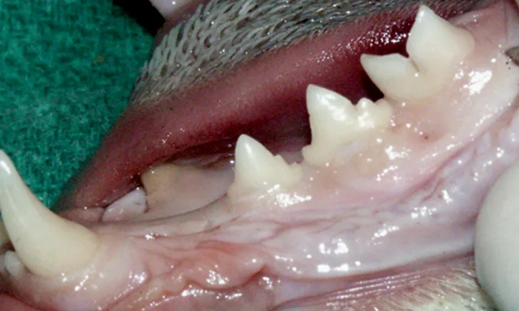
This image gallery demonstrates these techniques for extraction of incisor teeth, as well as canine, premolar, and molar teeth of the mandibular quadrant. Intraoperative views were obtained from a feline cadaver.

FIGURE 1A
A mucoperiosteal flap is created to extract teeth on the mandibular arcade. The incision line (A; dotted black line) is defined, then the following incisions are made: rostral vertical release (B; arrow) at the mesial aspect of the mandibular canine tooth, intrasulcar along the buccal aspect of the teeth and along the diastema between the canine and third premolar teeth (C), and caudal vertical release (D; arrow) at the distal aspect of the first molar tooth.