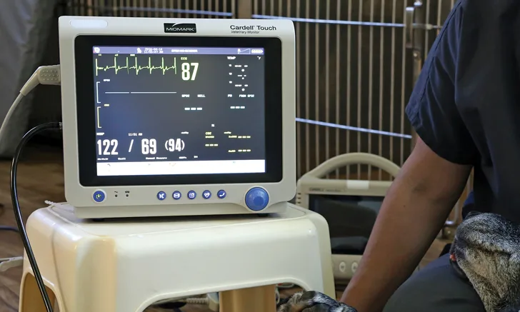
Clinical Cardiology History & Diagnostics
Wendy W. Mandese, DVM, University of Florida
Amara H. Estrada, DVM, DACVIM (Cardiology), University of Florida

For a comprehensive outline of the cardiology physical examination, see The Basic Cardiology Examination by Drs. Mandese and Estrada.
Recognizing subclinical heart disease in a healthy patient can be challenging. When heart disease is suspected, a patient may be referred to a board-certified cardiologist for diagnosis and treatment; however, specialist care may not be an option for all patients because of limited client finances, inconvenience of multiple veterinary appointments, and distance required to travel to a specialty practice. Primary care veterinarians should be proficient in performing cardiology evaluations and examinations to establish differential and definitive diagnoses in patients with cardiac conditions.
Valvular disease accounts for 75% to 80% of canine heart disease cases1 and is especially prevalent in small breeds. Myocardial disease is also common in dogs, with dilated cardiomyopathy occurring most often.2
In cats, more than 60% of heart disease cases are caused by hypertrophic cardiomyopathy.3 Dogs and cats may be presented with subclinical signs or emergently. Clinical signs vary depending on the species, disease process, and stage of cardiac disease.
Because clinical signs of cardiac disease can be subtle, obtaining a complete history is essential. The history should include specific questions about signs that the client might not associate with heart disease.
Signalment
Age
Clinical signs of cardiac disease in puppies and kittens may indicate a physiologic or congenital disease process. Puppies and kittens with a physiologic heart murmur do not show clinical signs of cardiac disease; the murmur in these patients is low grade (grade 1-2/6) and resolves before adulthood. The murmurs are usually systolic and loudest along the left sternal border.4
Breed
Many purebred dogs or cats are predisposed to specific congenital and/or acquired cardiac diseases (Tables 1 and 2). Certain breeds may benefit from screening (eg, auscultation, ECG, echocardiogram) during annual examinations. Clients should be educated about potential clinical signs.
Table 1: Common Breed Predilections for Congenital Cardiac Abnormalities5-9
History
Cough
In addition to cardiac disease, coughing can be a sign of another disease process. Coughing after eating or drinking suggests laryngeal dysfunction. Nocturnal coughs occur with cardiac insufficiency, pulmonary edema, and psychogenic conditions.10 If the cough is a dry hack or a goose-like honk, consider tracheitis, tracheal collapse, and compression of mainstem bronchi by left atrial enlargement or masses. Infectious tracheobronchitis or chronic bronchitis in dogs causes a productive cough and gagging. Cats with bronchial disease can experience episodic coughing, expiratory wheezes, and dyspnea. A soft, moist cough suggests pneumonia, parasitic or allergic disease, pulmonary thromboembolism, or edema.10
If coughing at home has been noted, the client should be asked what induces the cough (eg, exercise, excitement), about the quality of the cough (eg, dry, productive, moist), and how often the cough occurs. Clients often do not report coughing in cats but may report that the patient is gagging or retching, which sometimes progresses to vomiting. Thoracic radiographs should be obtained to determine if the cough is cardiogenic. Radiographic findings that indicate cardiomegaly (specifically, left atrial enlargement causing upward compression of the trachea and mainstem bronchi) may indicate a cardiac cause. In older, small-breed dogs, which are most commonly affected by congestive heart failure and airway disease (eg, tracheal collapse, chronic bronchitis), an airway cause for the cough should always be ruled out before diagnosing a cardiac cause. Pulmonary edema may be present radiographically in dogs with congestive heart failure but is unlikely to cause cough unless fluid accumulation is severe enough to build up in the airways.11
Activity Level
Cardiac disease can be an underlying cause of decreased activity and energy levels, and patients may have difficulty resting comfortably or may seem anxious and unable to relax.
Collapse Episodes
Syncope can be seen secondary to cardiac disease and is defined as a sudden, transient loss of consciousness originating from a short-term interruption of blood perfusion and oxygenation to the brain. It is characterized by sudden onset, short duration, and spontaneous recovery.12 Recovery can be so rapid that owners sometimes mistake the episodes for clumsiness or tripping. Collapse episodes can also be caused by neurologic incidents or seizures; determining whether collapse is caused by seizure activity or syncope can be difficult. In general, syncopal episodes are more often associated with excitement and exercise, and signs are less likely to occur leading up to the event. These episodes are usually of a shorter duration (1-2 seconds), and patients are also less likely to have signs following the event.12 Other causes of collapse episodes include anemia and orthopedic disease.
Appetite & Normal Behavior
A decrease in appetite or normal behavior, such as normal grooming behavior and desire to play or go for walks, can be an early sign of cardiac disease.
Table 2: Common Breed Predilections for Acquired Cardiac Abnormalities5-9
Diagnostics
Imaging
Radiography can be used to assess generalized cardiomegaly and chamber enlargement. Subjectivity, technique, and patient shape and size can limit the value of radiography, and obtaining diagnostic images can be challenging in cats, brachycephalic (ie, short-chested) dogs, and obese patients. Proper technique is important, as a film obtained on expiration or with the patient incorrectly positioned can result in an inaccurate cardiac silhouette and lead to misinterpretation.
Radiography is most useful for detecting sequelae to cardiac dysfunction, such as great vessel enlargement, pulmonary edema, and pleural effusion.13 It can also help rule out other causes of coughing (eg, primary pulmonary disease, collapsing trachea) and can be valuable in assessing changes consistent with heartworm disease in dogs.
Determining cardiac size can be subjective. The Vertebral Heart Scale (VHS) provides an objective method for measuring cardiac size in dogs. Starting with the cranial edge of the 4th thoracic vertebrae:

In dogs, normal VHS is 9.7 +/- 0.5 vertebral bodies.14 Studies have shown significant breed variation in VHS scores, with chondrodystrophic breeds having a greater VHS.15
An ECG should be performed in any patient in which an arrhythmia is detected, including bradycardia or sustained tachycardia. An ECG ideally should be performed in conjunction with echocardiography to determine the underlying cause of the arrhythmia.
Echocardiography should be used to assess chamber size and contractility of the heart and to obtain information about cardiac anatomy and function. Echocardiography is the most accurate diagnostic tool for most forms of heart disease.
Laboratory Studies
Baseline laboratory studies should be performed on all patients with suspected cardiac disease to identify other underlying disease processes that may affect diagnosis and treatment.
CBC
Increased turbulent blood flow resulting from decreased blood viscosity can be caused by anemia and results in a physiologic murmur. Increased leukocytes may indicate infections such as endocarditis and myocarditis; these diagnoses should be considered in patients that also have fever, marked lethargy, and a newly diagnosed murmur.
Serum Chemistry Profile
Chronic kidney disease can affect treatment options and prognosis in patients with heart disease. Increased liver enzyme values may be seen in patients with right-sided heart failure and in cats with hyperthyroidism.
Urinalysis
Urinalysis and urine culture should be performed to further assess renal function and evaluate for other underlying disease.
Thyroid Testing
In middle-aged and geriatric cats, hyper-thyroidism is a common cause of cardiac murmurs. Total T4 values should be obtained from cats in these age groups with a newly diagnosed murmur.
Heartworm Test
A heartworm antigen test should be conducted in any dog with suspected heart disease, especially if coughing is a clinical sign. Heartworm disease typically only causes a murmur in patients with caval syndrome, a condition in which worms present in the pulmonary artery ascend to the right ventricle and become intertwined with the tricuspid valve. Because testing for heartworms in cats can be diagnostically challenging, a combination of heartworm antibody and antigen tests is recommended.
Cardiac Biomarker Tests
Measurement of NT-proBNP and cardiac troponin can provide information about myocardial damage (eg, as in hypertrophic cardiomyopathy) and may help determine the significance of murmurs in cats. Patient-side snap tests for NT-proBNP have been helpful in differentiating between cardiac and noncardiac causes of respiratory distress in cats. The test can be performed using pleural fluid obtained by thoracocentesis, which may eliminate the need for blood testing and its associated stress.16