Placing a Central Sampling Catheter Through a Peripheral Vein
Kayla Hanson, DVM, Lakeshore Veterinary Specialists, Glendale, Wisconsin
Andrew Linklater, DVM, DACVECC, Veterinary Specialists of the Rockies, Castle Rock, Colorado
Critically ill patients often require frequent laboratory analysis for monitoring purposes but repeated venipuncture can lead to hematoma development, iatrogenic anemia, infection, and patient discomfort.1 A sampling catheter may be placed through a peripheral vein in hospitalized patients to facilitate repeated blood collection. In addition, access to the central (venous) circulation allows administration of hyperosmotic substances, parenteral nutrition, and measurement of central venous pressure.
If the catheter is used for repeated blood sample collection, this should be the catheter’s sole purpose. This is important to avoid sample errors that may result from administration of IV fluids and drugs through the same catheter lumen; some studies have shown that this may cause inaccurate diagnostic results, particularly with glucose, potassium, and venous blood gas analysis.2,3
A peripheral sampling catheter may be selected when frequent sampling is necessary, but a contraindication to true central venous catheterization via the jugular vein exists. Contraindications to jugular catheter placement include injury, infection, pain, or edema within the cervical region, coagulopathy, increased intracranial pressure, or cervical disk disease.
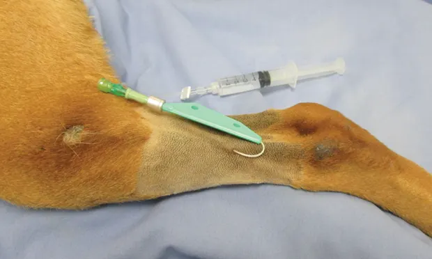
Lateral or medial saphenous veins are most commonly used for sampling catheter placement in canine and feline patients. Through-the-needle catheters are typically used, as they are small enough to pass through a peripheral vein and are longer (8–12 in) than over-the-needle catheters; this allows more central placement into the vessel and, in smaller patients, access to the caudal vena cava.
The catheters come in a variety of sizes and are appropriate for use in most patients (Table). As peripheral sampling catheters obtain closer proximity to the central circulation and are intended for longer term use, maintaining aseptic technique is important to decrease the risk of contamination, phlebitis, and catheter-associated infection or sepsis.
Table: Central Sampling Catheters Overview
*Lack of central venous access implies that (1) administration of hyperosmotic solutions should not be performed as it may result in phlebitis, (2) central venous pressures cannot be performed as an indirect assessment of intravascular volume, and (3) blood samples are difficult or impossible to obtain.
Thrombosis is a potential risk associated with long-term catheter use.4 Other disadvantages to this type of catheter include catheter shearing by the introducer needle, need for a bulky bandage to secure it in place, and small lumen size (limited by the needle), which may cause catheter occlusion. The potential for hemolysis exists when obtaining samples via these catheters (as opposed to venipuncture); however, recent studies of blood samples obtained via venipuncture versus through a peripheral catheter have shown no significant difference between the methods.5,6
Related Article: A New Technique for Emergency Vascular Access
Related Article: Placement of Central Venous Catheters
Catheter Management
Management of the catheter is important, to ensure patency and minimize catheter-related complications, such as phlebitis, infection, and thrombosis.4 The catheter should be flushed with 0.9% NaCl (with or without heparin) every 4–6 hours to ensure it remains patent. The bandage should be evaluated every 4–6 hours as well and replaced if soiled or wet. The bandage should be removed/replaced every 24–48 hours in as sterile a manner as possible so the limb and catheter insertion site can be evaluated. Evidence of phlebitis, infection, displacement, leakage, thrombosis, or a fever may prompt the clinician to remove the catheter.
Peripheral sampling catheters are a short-term option for vascular access; previous recommendations included catheter replacement every 72–96 hours, however, because of limited peripheral venous access sites and lack of evidence to support such frequent replacements, recent recommendations suggest that as long as appropriate catheter care is performed and no complications noted, peripheral catheters need only be replaced when clinically indicated, rather than on a scheduled basis.7
Step-by-Step: Placing a Central Sampling Catheter Through a Peripheral Vein
What You Will Need
Clippers
5% chlorhexidine scrub
Sterile drape
Sterile gloves
Through-the-needle catheter kit
Injection cap or T-set
Saline flush
1-inch tape
Bandage material

STEP 1
Place the patient in lateral recumbency and clip hair around the saphenous vein, allowing margins of at least 2 cm. Sedation may be necessary as dictated by patient needs.
Author Insight
Take care when placing a through-the-needle long catheter in a coagulopathic patient, as excessive bleeding may occur. This is easily limited, however, with digital or bandage pressure.