How to Assess Phalangeal Fractures
Rachel Basa, BVSc, DECVS, GradCertVS, MANZCVS, MVCS, PhD, The University of Sydney, Camperdown, Australia
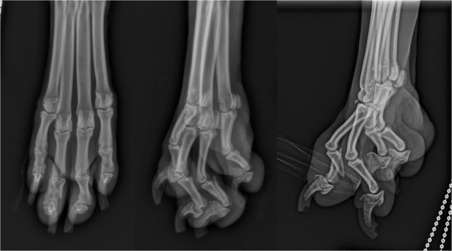
Phalangeal fractures are a relatively common cause of lameness in cats and dogs, but literature describing incidence and management are sparse. In an early report, phalangeal fractures accounted for 2.8% of all fractures in dogs and 0.7% in cats.1 In the author’s experience, phalangeal fractures are frequently missed at primary care clinics due to superimposition of overlapping bones and inadequate collimation to the region of interest. Left untreated, these injuries can result in persistent, chronic lameness due to malunion or nonunion, secondary nail bed injuries, and osteoarthritis or chronic instability if the collateral ligament insertions are affected.2
Phalangeal Anatomy
The digit consists of 3 bones, including proximal, middle, and distal phalanges in addition to 2 large palmar sesamoid bones. The middle phalanx is present in each of the digits, except for digit 1 of the carpus.3 Ligamentous stabilizers of the phalanges include collateral ligaments of the metacarpophalangeal and metatarsophalangeal joints and the proximal and distal interphalangeal joints (Figure 1). An additional 4 stabilizing ligaments (cruciate and sesamoidean) are at the level of the metacarpophalangeal and metatarsophalangeal joints.3 The proximal and middle phalanges have a base, head, and body.
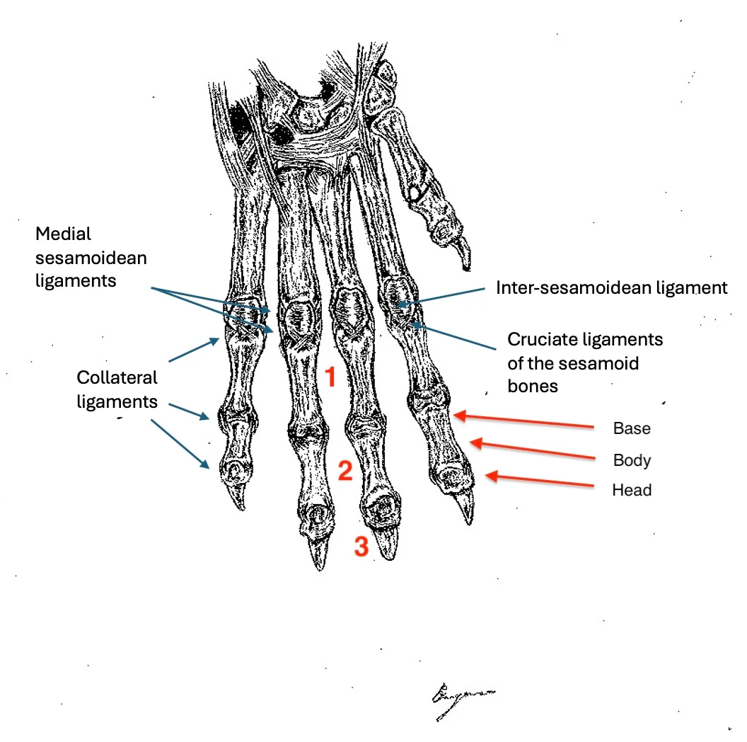
FIGURE 1 Ligaments of the phalanges; base (1), body (2), and head (3) of the middle phalanx of digit II, corresponding to the proximal, middle, and distal phalanges, respectively. Image illustrated by Elizabeth Street.
Common fracture configurations that may be identified in association with the proximal and middle phalanges include transverse, long oblique, and spiral extra-articular fractures of the body. Comminuted fractures that are either extra-articular or extend to the joint at the base or head of the affected phalanx are also possible.4
Presentation
Cats and dogs with phalangeal fractures are typically presented with non–weight-bearing lameness and swelling, pain, and/or crepitus on palpation of the affected digit. Lameness may be acute or chronic. The most common etiology is trauma (eg, crush injuries, high impact athletic activity, bite wounds). On examination of the digit, the affected phalanx should be isolated, and the integrity of the collateral ligaments can be assessed by applying varus and valgus stress. Sedation should be considered for patients with injuries that are too painful to allow complete orthopedic examination.
Diagnostics
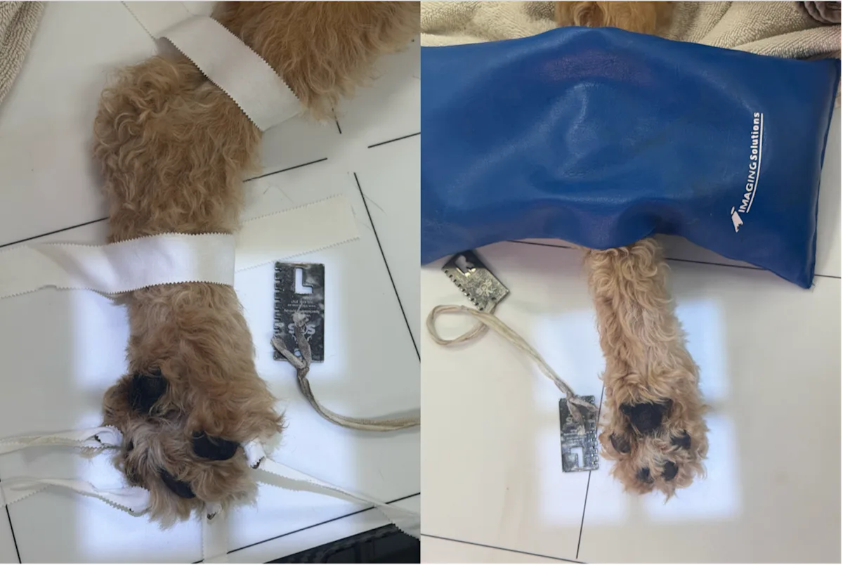
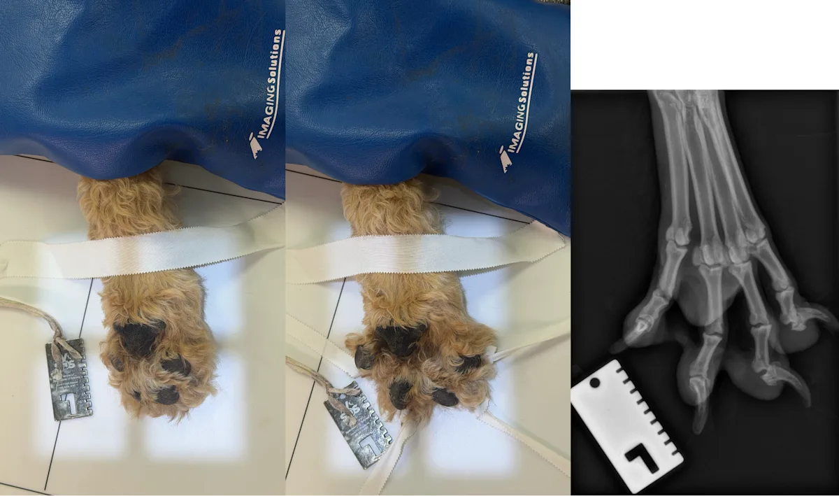
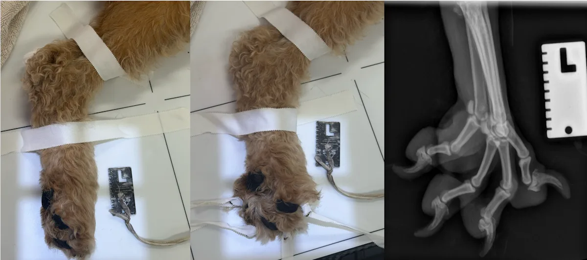
Fractures are often detectable on plain radiographs, with dorsopalmar/dorsoplantar (DP) and lateral views required at minimum to assess for phalangeal injuries. Additional DP 15-degree oblique views and lateral 45-degree oblique views can assist in the isolation of individual bones (Figures 2 and 3).5 Tape can be used to splay the digits for lateral and DP views.5 In cases in which the fracture cannot be identified clearly on plain radiographs, CT can be useful to characterize complex fractures and facilitate surgical planning (Figure 4).
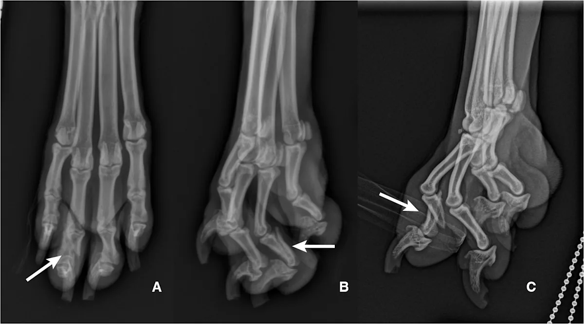
FIGURE 2 Dorsoplantar (A) and lateral (B) radiographs of the right pes of a 3-year-old neutered male mastiff crossbreed with a 2-week history of right pelvic limb lameness. The lateral oblique view (C) demonstrates a short oblique fracture affecting P2 of digit 3.
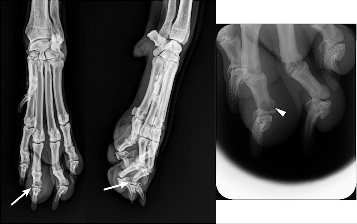
FIGURE 3 Dorsopalmar (left) and lateral (right) radiographs of a 3-year-old spayed border collie with a 2-month history of intermittent non–weight-bearing left thoracic limb lameness. Pain and swelling were associated with P2 of digit 3. An area of radiolucency associated with the distal aspect of P2 can be seen on the dorsopalmar radiograph (arrows) and on a further collimated view (arrowhead).
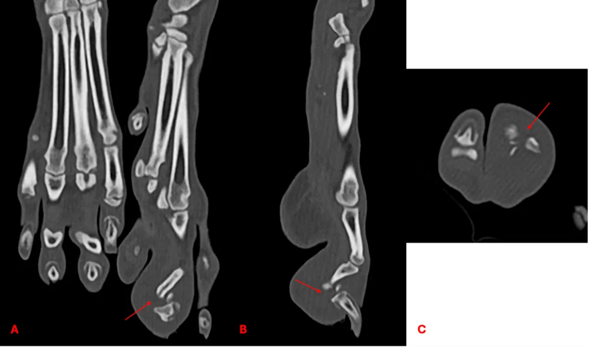
FIGURE 4 CT of the patient in Figure 3 with multiplanar reconstruction (A, frontal plane; B, sagittal plane; C, transverse plane). Comminuted articular fracture of the distal epiphysis of P2, digit 3 (arrows) was confirmed.
Treatment
Treatment options for phalangeal fractures include conservative management with or without external coaptation, internal fixation (with bone plates, lag screws, dowel pinning), excision arthroplasty of the interphalangeal joint, and arthrodesis.4-6 Conservative management with external coaptation can be recommended in patients with closed, nondisplaced, extra-articular fractures and fractures affecting digits 2 and 5 and may result in a fibrous union that can lead to return of normal limb function. Buddy taping entails encompassing the fractured and adjacent intact digits in elasticized tape that is 1 inch in width.2,5 Following tape application, a soft padded bandage can be applied from the distal tibia or radius to cover the digits until the bone is healed (3-4 weeks).5 If this splinting technique is not used, a custom palmar or plantar splint can be applied with a soft padded bandage for 6 weeks. During this time, the author recommends crate confinement and repeat radiography after 6 to 8 weeks.
Surgical stabilization is indicated with fracture displacement >50%, inability to reduce the fracture, high-performance athletic patients, or intra-articular or open fractures. In some cases, surgical stabilization may not be possible due to the small size of the bones. Digit amputation may be considered. Short- and long-term complications (eg, incisional dehiscence, lameness) of digit amputations are reasonably high (39% and 25%, respectively, in a case series),7 but 95.8% of pet owners indicated satisfaction with surgery results.7 Amputation should also be considered for any open or comminuted digit fracture or articular fracture of the distal phalanx.2 In high-performance athletic patients, excision arthroplasty or arthrodesis may be preferred instead of amputation.
Step-by-Step: Palpation & Radiography of Phalangeal Fractures
What You Will Need
Adhesive tape
Radiographic positioning aids, including bean bags and a U-trough
Radiography imaging unit
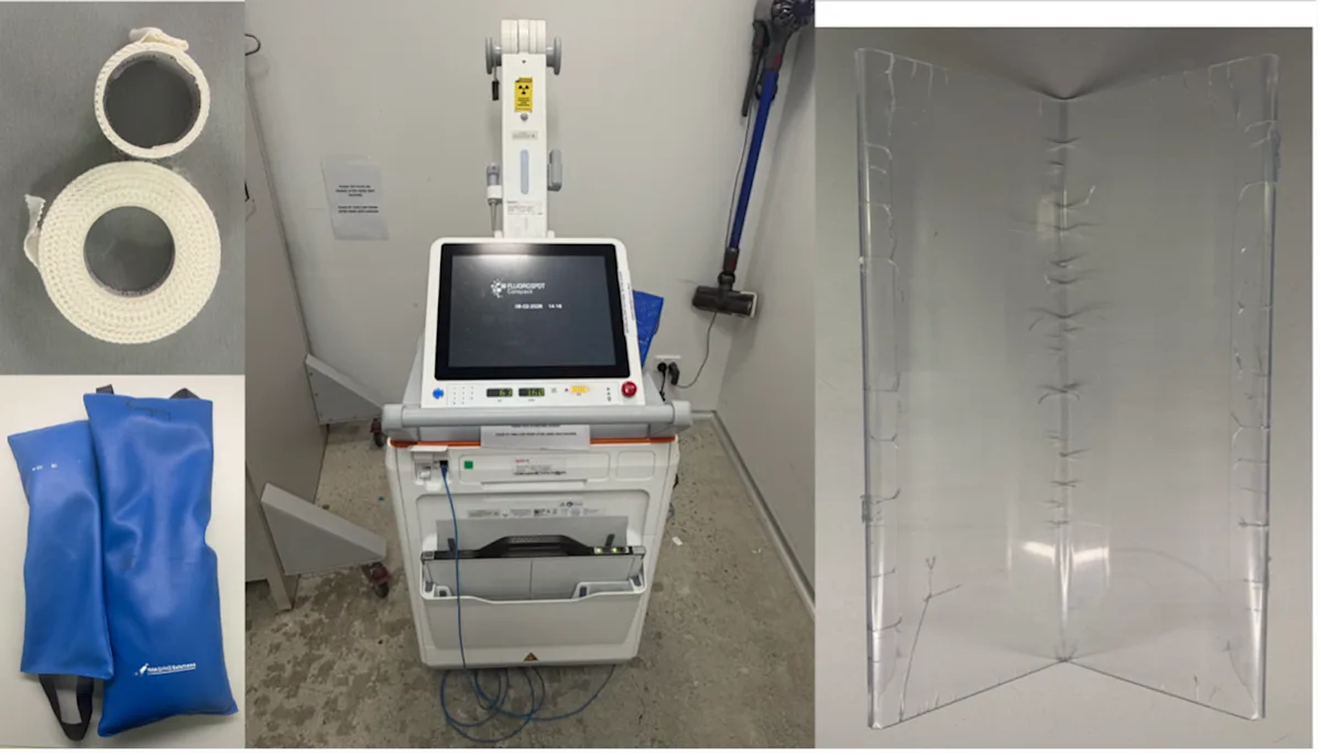
Step 1
Following a complete orthopedic examination, sedate the patient, and palpate each digit and individual phalanx for evidence of swelling, crepitus, and/or pain.
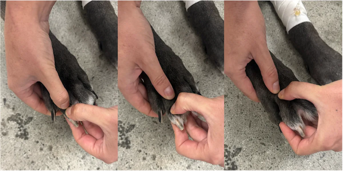
Step 2
Test for varus and valgus instability of each phalanx.
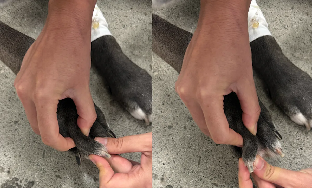
Step 3
Perform plain orthogonal radiography of the affected manus/pes. Collimate to the region of interest.
Step 4
Position the patient for additional DP 15-degree oblique views (top) and lateral 45-degree oblique views (bottom) to enable isolation of individual digits. Use tape if needed to isolate individual digits.
Author Insight
If phalangeal fracture is suspected despite a lack of obvious abnormalities on radiographs, referral for a CT scan should be considered.