Nutritional Support for Critically Ill Dogs & Cats
Lisa P. Weeth, DVM, MRCVS, DACVN, Weeth Nutrition Services, Los Angeles, California; Gulf Coast Veterinary Telemedicine, Houston, Texas
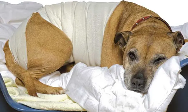
Nutritional support for critically ill dogs and cats is often delayed until diagnosis or appropriate medical/surgical intervention can occur.
Anorexia is a lack of food intake. Hyporexia is a reduction in food intake. Lack of food intake may not be recognized until cachexia or biochemical evidence of starvation occurs.
A 2001 survey found that, in hospitalized dogs, 73% of the days admitted were spent in negative energy balance. Reasons cited were poorly written orders (22%), orders to withhold food (34%), and the animal’s refusal to eat (44%).1
Animals in negative energy balance may be protein–calorie malnourished and have whole-body depletion of essential nutrients.
Pathophysiology
Animals undergoing simple starvation should be differentiated from those with medical conditions that result in physiologic stress or impaired hormone balance. Withholding energy and key nutrients from critically ill patients may cloud diagnosis, hinder response to treatment, and increase risk for sepsis.
Protein–calorie malnutrition results in decreased immune competence and catabolism of not only lean body mass, but organ and lymphatic tissues.
B vitamins are necessary for energy metabolism and adequate lymphocyte function.
Glutamine is important for normal enterocyte health; arginine and taurine are important for lymphocyte function.
Deficiencies in certain minerals, such as selenium, copper, zinc, and iron, increase rates of infection and decrease cell-mediated and humoral responses.
Deficiencies in vitamins A and E result in impaired lymphocyte function and delayed wound healing.
Causes & Risk Factors
Animals living in multianimal households or that have several caregivers may have prolonged hyporexia that goes unnoticed for days to weeks.
Overweight or obese animals may have significant muscle wasting that is overlooked due to degree of adiposity.
Animals on unconventional diets (eg, unbalanced home-prepared diets) may have whole-body depletion of essential nutrients that may impact immune function, wound healing, and response to medications.
Clinical Signs
Clinical signs of anorexia and hyporexia are often evident during the initial physical examination:
Body condition score—using a 9-point or 5-point scale—should be evaluated on initial presentation.
Muscle catabolism is often evident along the epaxial muscles; evaluation of lean muscle mass should be performed daily on hospitalized patients.
Weight change often reflects alteration in water balance and is a poor indicator of nutritional status.
Diagnostic Tests
There are no specific diagnostic tests to evaluate nutritional status. Serum albumin, mineral, and electrolyte levels can be affected by multiple factors. Diagnostic tests should be directed at determining the underlying cause of illness.
History
Diet, consumption of treats, and supplement history will help direct feeding plans. This step is not essential for initial stabilization and treatment of the acutely ill animal, but it helps in developing an appropriate feeding and treatment plan.
Duration of acceptable starvation may differ for animals with simple starvation due to acute trauma or illness versus those with complex medical conditions.
Animals with an ideal BCS and adequate lean muscle mass may require approaches that differ from those for obese animals with palpable muscle wasting.
Animals with fixed flavor or texture preferences (especially cats) will continue to refuse foods with an assumed high palatability due to learned food preferences.
Treatment
Indications for nutritional support include:
Anorexia > 3 days; expected anorexia > 3 days
History of hyporexia > 7 days
Weight loss > 10% body weight
Expected ongoing nutrient loss, high energy demands, or hypoalbuminemia.
Treatment at a Glance
Identify the cause of anorexia/hyporexia
Calculate RER: 70 × BWkg0.75
Select the feeding method:
Oral: Voluntary or prompt through pharmacologic agent
Assisted enteral: Nasoesophageal/nasogastric, esophagostomy, gastrostomy, or jejunostomy
Parenteral
Start with 25% RER and increase to meet full RER over 2 to 4 days
Monitor response to refeeding:
Body weight
Lean muscle mass
Serum electrolytes
Adjust calories and nutrients delivered as needed
Energy Requirements
While in the hospital, feed to meet the resting energy requirement (RER), calculated according to one of the following formulas, and adjust as needed.
Allometric formula (preferred; can be used for all animals)
RER = 70 × BWkg0.75
Linear formula (can be used for animals between 2 and 35 kg)
RER = [30 × BWkg] + 70
Avoid overfeeding hospitalized dogs and cats—providing excess calories can result in hyperglycemia (if given parenterally) or regurgitation, abdominal cramping, vomiting, and diarrhea (if given enterally). Hyperglycemia has been shown to negatively impact recovery and subsequent discharge from the hospital; if vomiting or regurgitation occurs, there is an increased risk for aspiration.
Selecting the Feeding Method
Planning should begin on the first day of hospital admission.
Selection should be based on patient signalment, BCS, disease state, expected duration of inadequate intake, function of the gastrointestinal tract, and the patient’s ability to tolerate anesthesia.
Voluntary Food Intake
Avoid or minimize negative environmental hindrances to food intake, including noise, frequent disturbances, bowls near litter boxes (cats), and electronic collars.
Do not offer several diets at one time. This may result in learned food aversion.
Do not force-feed ill animals. This can exacerbate food aversions and may cause aspiration.
Pharmacologic intervention may be considered if short-term intervention is needed; however, animals will not consume full RER with appetite stimulants alone.
Cyproheptadine
Megestrol acetate
Diazepam
Nasoesophageal or Nasogastric Tubes
Indications: Short-term (< 10 days) in the hospital; indicated in animals with normal nasal, pharyngeal, esophageal, and gastric function that are unable to be placed under anesthesia. Little to no anesthesia is required.
Particulars:
Narrow-diameter tubes (5- to 8-Fr)
Liquid enteral solution required due to tube-size limitations. Nutrient profiles of some solutions may not be suitable for animals with certain diseases.
Feedings are best delivered as a constant rate infusion to minimize risk for regurgitation and aspiration.
Tubes placed into the stomach can also be used to relieve air trapped in the stomach.
Complications: Epistaxis, facial irritation, and premature tube removal are common complications.
Step by step guide: Nasoesophageal or Nasogastric Feeding Tube Placement
Esophagostomy Tubes
Indications: Placed in the hospital; maintained at home for weeks to months as needed. General anesthesia required, but placement is relatively quick and easy.
Particulars:
Larger diameter tubes (12- to 14-Fr for cats and dogs weighing < 15 kg; 14- to 18-Fr for dogs > 15 kg)
Most commercial canned diets can be fed; blend with water.
To ensure adequate delivery, energy density of final slurry (divide final volume by number of calories contained therein) must be calculated. It should be close to 1 kcal/mL.
Well tolerated by most animals; can be used for medication administration at home even after the animal resumes normal food intake.
Complications: Minimal risk for life-threatening complications if tubes removed early; most common complication is stomal site infection.
Step by step guide: Esophagostomy Feeding Tubes
Gastrostomy Tubes
Indications: Animals requiring long-term nutritional support (months to year) or those with esophageal pathology (esophagitis, megaesophagus, or esophageal strictures). Can be placed endoscopically (percutaneous endoscopic gastrostomy), via percutaneous blind placement, or surgically; dogs > 20 kg or those that are obese require surgical placement.
Particulars:
Larger diameter tubes (18- to 24-Fr)
Wide range of food selections; blend with water
To ensure adequate delivery, calculate energy density of final slurry (see calculation previously mentioned). Energy density should be > 1 kcal/mL.
Requires longer anesthesia +/- specialized equipment to place
Low-profile gastrostomy tubes well tolerated
Complications: Life-threatening complications (peritonitis, gastric hemorrhage) if tube removed prematurely
Jejunostomy Tubes
Indications: Animals unable to tolerate gastric feeding that have normal jejunal, ileal, and colonic function. May be indicated in animals with gastric outflow or proximal small intestine obstructions or severe pancreatitis. Requires general anesthesia; best used in a veterinary setting where continued monitoring and care can be provided.
Particulars:
Surgical placement most common but newer percutaneous endoscopic gastrojejunostomy tube placement techniques have been described.
Narrow-diameter tubes (5- to 8-Fr)
Liquid enteral solution required due to tube size limitations. Lower-fat human monomeric liquid solutions (eg, Vivonex Plus; nestle-nutrition.com) may be required for animals requiring fat restriction.
Solutions are delivered as a constant-rate infusion over 12 to 16 hours to prevent complications, such as abdominal cramping and diarrhea.
Complications: Include peritonitis if tube removed prematurely
Parenteral Nutritional Support
Indications: Animals with protracted vomiting, severe pancreatitis, extensive gastrointestinal tract disease, and weak-to-absent gag reflexes; animals receiving mechanical ventilation; and animals that are obtunded or unable to maintain sternal recumbency.
Particulars:
Total parenteral nutrition (TPN) must be delivered through a dedicated central catheter. Solutions should be compounded for the individual animal.
Peripheral parenteral nutrition (PPN) can be delivered through a dedicated peripheral catheter. Individually compounded or commercially available PPN solutions can be used.
Catheters and fluid lines must be handled aseptically and disconnected only to change the parenteral solution bag. Animals should be monitored closely in the hospital for biochemical disturbances.
Follow-Up
Patient Monitoring
Like any treatment plan, nutritional support should be dynamic. Start with a goal to deliver 25% of calculated RER on day 1, and increase gradually until full RER is reached in 2 to 4 days. Adjust the amount delivered every 12 to 24 hours based on body weight changes, physical examination findings, and known or expected ongoing losses.
If the patient is receiving assisted enteral feeding, deliver food slowly to avoid vomiting, regurgitation, and abdominal cramping.
Initial enteral feedings should provide 5 to 10 mL/kg per feeding and can be increased to 10 to 20 mL/kg in many animals.
Measure and record body weight daily.
Check electrolytes 24 hours after starting to monitor for hypokalemia, hypophosphatemia, and hypomagnesemia.
Relative Cost
The cost of nutritional support varies according to the underlying disease state, feeding method, and monitoring. Total cost of diagnosis, hospitalization, treatment, and assisted nutritional support will often reach thousands of dollars, depending on disease state and severity.
Prognosis
The outcome for critically ill animals depends on the nature of anorexia or hyporexia, existing comorbidities, and duration of cachexia before intervention. There is evidence that early enteral nutritional intervention may shorten recovery time and minimize bacterial translocation across the gut.