In the Literature
Baldeon M, Perry KL. Medial humeral epicondylitis: a retrospective case series of nine cats (17 elbows). J Feline Med Surg. 2025;27(7):1098612X251347952. doi:10.1177/1098612X251347952
The Research …
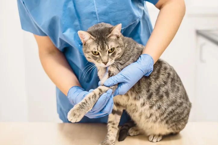
Medial humeral epicondylitis (MHE) is an underdiagnosed cause of thoracic limb lameness in cats that is associated with degeneration and mineralization of the flexor carpi ulnaris tendon at the medial humeral epicondyle. Data describing clinical features, diagnostic findings, and treatment outcomes in affected cats are limited.
This retrospective study reviewed records of 9 cats (17 affected elbows) radiographically diagnosed with MHE. All cats underwent initial nonsurgical management, with a subset later requiring surgical intervention. Diagnostic workup included radiography of all elbows, CT of 10 elbows, and ultrasonography of 4 elbows.
Median age and weight at time of diagnosis were 6 years (range, 1-15 years) and 15.2 lb (range, 7.3-18 lb; 6.9 kg [range, 3.3-8.2 kg]), respectively. Clinical presentation included insidious moderate lameness, pain on palpation caudodistal to the medial epicondyle, pain with carpal flexion, and pain with elbow pronation or supination. Palpable thickening and mineralization distal to the medial epicondyle were also noted in multiple cats.
Radiographically, epicondylitis was graded as mild (n = 8), moderate (n = 7), or severe (n = 2). In 2 cases (4 elbows) with mild or equivocal radiographic findings, early changes (ie, fluid, enthesopathy, small focal mineralization) were detected on ultrasonography. CT showed additional findings in 7 elbows (including intra-articular mineralized bodies in 5 elbows) that influenced surgical planning in some cases.
Nonsurgical management (ie, NSAIDs, analgesics, activity modification, physical rehabilitation) resulted in full resolution in 1 cat, partial resolution in 4 cats, and no improvement in 4 cats. Better responses were observed in cases with mild radiographic changes. Surgical treatment in 4 cats (7 elbows) resulted in complete resolution in 3 cats (5 elbows); 1 cat had improved but residual lameness attributed to concurrent elbow osteoarthritis.
… The Takeaways
Key pearls to put into practice:
MHE should be suspected in all cats presented with insidious thoracic limb lameness with pain over the medial epicondyle, especially if there is no history of trauma, even in indoor and young cats. Unlike previous reports,1,2 in which most cats were older with outdoor access, all cats in this study were housed indoors without outdoor access.
Ultrasonography enables early detection of fluid, enthesopathy, and small mineralization before advanced radiographic changes appear. Radiography better detects advanced disease, having low sensitivity in early cases. In this study, CT identified intra-articular bodies in >50% of elbows.
Nonsurgical management of MHE (eg, NSAIDs, rest, weight management, restriction of jumping, physical therapy) may be attempted initially, especially if radiographic changes are mild; however, response is limited in moderate to severe or chronic cases. In cats with lack of improvement after 2 to 4 weeks or evidence of moderate to severe changes, surgery is likely indicated.
Surgical procedures include removal of mineralized tissue from the flexor carpi ulnaris, loosening or releasing the ulnar nerve from surrounding scar tissue to improve nerve function, and, in some cases, arthrotomy to remove intra-articular bodies. Postsurgical prognosis is generally good. When combined, the results of this study and another with clinical follow-up showed 10 out of 13 cats were free of clinical lameness 8 to 12 weeks after surgery.2
MHE is likely underdiagnosed. Based on a postmortem study, ≈10% of cats were affected by MHE.1
You are reading 2-Minute Takeaways, a research summary resource presented by Clinician’s Brief. Clinician’s Brief does not conduct primary research.