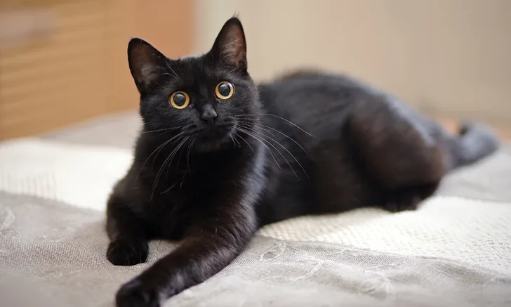
Lower Urinary Tract Signs in a Cat
Joe Bartges, DVM, PhD, DACVIM (SAIM), DACVN, Cornell University Veterinary Specialists
Kara M. Burns, MS, MEd, LVT, VTS (Nutrition), Olathe, Kansas
Gregg K. Takashima, DVM, WSAVA Global Nutrition Committee Series Editor

The Case
Inky, a 7-year-old, 7.8-lb (3.55-kg), neutered male domestic shorthair cat, was presented for recurrent signs of lower urinary tract disease (LUTD) characterized by urinating outside the litter box, hematuria, and pollakiuria.
History disclosed at least 5 episodes of LUTD over 2 years. Urinalyses, when performed, had revealed only hematuria; crystalluria and pyuria were not present. Other parameters on CBC and serum chemistry profile were normal. One urine culture had been performed and revealed no growth. Inky had previously been treated with amoxicillin, amoxicillin–clavulanic acid, and prednisolone, and his diet was changed after the second LUTD presentation. Clinical signs for each episode were reported to have resolved after therapeutic intervention over 4 to 14 days.
Examination
Physical examination showed the patient was bright and alert with a palpable, moderately distended urinary bladder that was not painful. Blood work (ie, CBC, serum chemistry profile, total thyroxine) results were within normal limits. Urinalysis via cystocentesis showed a urine specific gravity (USG) of 1.045 and a pH of 6. RBCs per high-power field were too numerous to count. Other results were within normal limits. Urine culture showed no growth after 72 hours. Muscle mass was normal, and BCS was 5 of 9.
Dietary History
A comprehensive nutritional evaluation was completed. The patient had been eating a therapeutic canned struvite and oxalate preventive diet for approximately one year. He was occasionally (<1 time/week) fed over-the-counter treats. He was an indoor-only cat in a household with no other pets.
Related Article: Managing Indwelling Urinary Catheters
Related Article: Urinary Catheter Placement for Feline Urethral Obstruction
Testing & Treatment
Abdominal radiography and ultrasonography were conducted. Abdominal radiography revealed no abnormalities (Figure 1); ultrasonography revealed 2 small urocystoliths (Figure 2). Medical dissolution was not attempted because the uroliths were not believed to be composed of struvite due to their radiolucency, lack of crystalluria, and aciduria. Because the patient was a male cat, no attempt was made to retrieve the urolith by voiding urohydropropulsion. Percutaneous cystolithotomy1 (Figure 3) was performed. Urocystoliths (Figure 4) were removed and submitted for quantitative analysis.

FIGURE 1
Lateral abdominal radiograph of patient
Diagnosis: Ammonium Urate Urocystoliths
Idiopathic Urate Urolithiasis
Ammonium urate accounted for approximately 5% of uroliths obtained from cats and submitted to the Minnesota Urolith Center in 2016.2 Urate uroliths form as a result of liver disease (usually a portosystemic vascular shunt)3,4 or because of an inborn metabolism error that causes hyperuricosuria (eg, in dalmatians and English bulldogs).5,6 However, the underlying mechanism for ammonium urate urolithiasis in cats without liver disease is unknown. Idiopathic urate urolithiasis usually occurs in cats between 4 and 7 years of age5-7 but can occur in kittens secondary to congenital liver disease (eg, portovascular anomalies).8
There are no published protocols for medical dissolution of ammonium urate uroliths in cats with idiopathic urate urolithiasis.
Follow-Up
Despite normal laboratory results, provocative bile acid testing was performed; results were normal.
Nutritional Consultation
In the author’s experience, prevention of urate urolith recurrence is successful in more than 90% of feline cases in which a therapeutic kidney diet is fed.9 There are several goals of nutritional treatment of urate urolithiasis in cats. The diet should be low in purines; however, the purine content of a diet is not provided on food labels. Thus, a low-protein diet is often fed (<30% on a dry matter basis). In addition, feeding a canned product to induce more dilute urine (USG <1.030) is preferred over feeding a dry product. The diet should also induce alkaluria (urine pH >7).
Inky was transitioned to a canned therapeutic kidney diet with instructions to minimize treats. Periodic monitoring via urinalysis and abdominal ultrasonography, as well as follow-up with the owner and patient by the nutritional healthcare team, has shown no recurrence in more than 5 years.
Conclusion
This case illustrates that not all cats with LUTD have idiopathic cystitis or uroliths composed of struvite or calcium oxalate.10,11 Thorough evaluation of a patient, including an in-depth nutritional history, is important, especially when clinical signs are recurrent or when response to treatment does not match expectations.
ASK YOURSELF …
LUTD = lower urinary tract disease, USG = urine specific gravity
Diet in Disease is a series developed by the World Small Animal Veterinary Association (WSAVA), the Academy of Veterinary Nutrition Technicians, and Clinician’s Brief.
Editor's Note: The author is an advisory board member for Blue Buffalo and a consultant for Veterinary Information Network.