Urinary Catheter Placement for Feline Urethral Obstruction
Garret E. Pachtinger, VMD, DACVECC, Veterinary Specialty and Emergency Center, Levittown, Pennsylvania
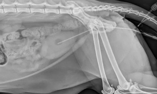
Clinically, urethral obstructions typically occur because of a mucus plug, calculus, or stricture. Other causes include a mechanical obstruction secondary to urethral spasm or a neoplastic process. While both male and female cats may develop clinical signs consistent with lower urinary tract disease (eg, stranguria, hematuria, pollakiuria), the male urethra is narrower, longer, and predisposed to obstruction.
Pathophysiology
The most common cause of urethral obstruction in cats is an underlying idiopathic sterile cystitis. Idiopathic cystitis may result from a neurohumoral alteration, notably an imbalance between the sympathetic nervous system and the hypothalamicpituitaryadrenal axis, causing blood flow alterations, increased circulation of inflammatory mediators and subsequent edema, smooth muscle spasm, and discomfort. Ultimately, the combination of inflammation, a mucus plug, and urethral spasm can result in urethral obstruction.
In addition to relieving the obstruction, life-threatening sequelae must be addressed. In complete urethral obstruction, the bladder distends until increased internal pressure results in necrosis and mucosal injury. This pressure is transmitted to the ureters and subsequently to the kidneys, exceeding the glomerular filtration pressure and resulting in azotemia, electrolyte derangements (eg, hyperphosphatemia, hyperkalemia), acidosis, and decreased urine production.
History & Clinical Signs
Patient history may include vocalization, stranguria, pollakiuria, hematuria, periuria, possible constipation, lethargy, excessive grooming of the perineal region, increased litter box visits, vomiting, and/or inappetence. Clinical signs vary depending on duration of obstruction. Patients presented early in the disease process may be clinically normal, aside from a firm, inexpressible urinary bladder that may be painful on palpation. Dehydration is also common; the patient is often anorexic, not drinking, and has ongoing losses (eg, vomiting). Signs are likely to occur when the obstruction has a longer duration (>24 hours).
More severe systemic signs may include weakness, collapse, and cardiac arrhythmia. Hyperkalemia, common in cats with prolonged obstruction, affects the electrical conduction through the heart by raising the resting membrane potential of myocytes. As the resting potential becomes less negative, sodium channels are affected, resulting in bradycardia. Severe hyperkalemia may result in bradycardia, sinoatrial arrest, and the potential for asystole.
Related Article: Urinary Obstruction: Treatment Measures
Related Article: Emergency Management of Urethral Obstruction in Male Cats
Initial Assessment & Diagnostics
Initial assessment and stabilization are often achieved simultaneously with IV catheter placement. To reduce the need for repeated phlebotomy, blood samples are obtained during catheter placement. Initial blood work should include PCV/TP, blood glucose, creatinine, venous blood gas analysis, and electrolytes. CBC and serum chemistry panel should also be conducted later.
When a patient is presented with suspected urethral obstruction, initial actions should include
Checking for dehydration, cardiovascular shock, bradycardia, and hypothermia
Performing an ECG
Measuring electrolytes and acid-base status and evaluating azotemia
Classic ECG changes associated with hyperkalemia include prolonged P-R interval, absent P waves, widened QRS complex, and tall or tented T waves.
Emergency Stabilization
After catheter placement and phlebotomy, isotonic crystalloid fluid therapy (eg, lactated Ringer's solution, 0.9% NaCl) is often started to address common abnormalities (eg, dehydration, hypotension, electrolyte derangements).
Despite concern about IV fluids distending the bladder further (with the potential for rupture), this rarely occurs. The pressure transmitted from the bladder to the kidneys reduces the glomerular filtration rate and decreases urine production, so the bladder is not often impacted in this early phase of treatment.
Unless the patient is unstable, urethral catheter placement is impossible or the bladder is distended to the point that the clinician feels uncomfortable flushing during unblocking, cystocentesis should be avoided. There is considerable risk for rupture if the large, distended, firm bladder–already at risk for pressure necrosis or mucosal injury–is pierced.
Although urinary catheter placement to relieve obstruction and IV fluid administration ultimately reverse the adverse effects associated with hyperkalemia, uremia, and acidosis, these treatments alone do not result in rapid effects. If the patient exhibits severe complications from hyperkalemia, particularly sinoatrial arrest and bradycardia (heart rate, <140 bpm), immediate intervention to protect the heart from hyperkalemia should be implemented (see Interventions to Protect the Heart).
Interventions to Protect the Heart
Calcium gluconate
A dose of 50–100 mg/kg IV administered slowly (over 10–20 min) will antagonize the cardiac effects of hyperkalemia.
The effects are immediate, lasting ~20–30 min.
Regular insulin or dextrose
A dose of 0.1–0.25 U/kg of regular insulin IV can be used to promote an intracellular shift of potassium. It takes about 20–30 min following administration to see an effect.
To prevent hypoglycemia, insulin administration is followed by a glucose bolus of 1–2 g per unit of insulin given.
Following insulin administration, blood glucose is monitored q2–4h for 6–12 hours with glucose supplementation as needed to maintain normoglycemia.
Sodium bicarbonate
Sodium bicarbonate lowers plasma potassium concentration by raising the blood pH and driving potassium intracellularly.
The formula used to calculate the MEq of sodium bicarbonate is:
0.3 body weight (kg) x base units in MEq/L deficit
This gives an approximation of the total bicarbonate deficit. One-third to one fourth of this dose is administered slowly by IV followed by patient and blood work reassessment to determine response and whether additional dosing is needed.
It usually takes 20–30 min to see an effect with this medication.
Catheter Placement
This life-threatening illness is ultimately treated by placing a urinary catheter to relieve obstruction. The effects on each patient must be evaluated individually to determine the degree of chemical restraint required. In severely azotemic, hyperkalemic, and obtunded patients, limited chemical restraint may be required.
While general anesthesia can be considered, the author commonly uses a combination of ketamine, diazepam, and butorphanol (or buprenorphine) for heavy sedation along with supplemental oxygen via flow-by or mask placement. For patients in which ketamine is less desirable (eg, heart disease), propofol can be substituted. After the patient has been sedated or anesthetized, urinary catheter placement can begin.
Postprocedural Care
Most patients remain hospitalized for 36 to 48 hours for continued monitoring (including urine volume), balanced fluid therapy, and assessment of urine character.
Patients with more serious illness or azotemia are also monitored for postobstructive diuresis, a clinical syndrome leading to significant urine production resulting from osmotic diuresis and medullary washout. These patients must be monitored closely, as their urine volume may be as high as 100 to 150 mL/hr. Some cats require high fluid rates to match the significant urine production.
Other important postprocedural treatment goals include pain management (buprenorphine, 10–15 g/kg IV q4–6h or butorphanol, 0.1–0.4 mg/kg IV q4–6h), urethral spasm treatment (phenoxybenzamine, 0.25–0.5 mg/kg PO q12–24h; prazosin, 0.25–1 mg PO q8–12h), and electrolyte monitoring. Electrolytes should be monitored q12–24h, or more often if significant abnormalities exist. Hyperkalemia may be noted on presentation, but hypokalemia may be seen after obstruction relief and postobstructive diuresis.
Antibiotics are often unnecessary; few cats have a true bacterial urinary tract infection on presentation, and inappropriate antibiotic use may result in a drug-resistant infection. Urine culture should be submitted if the urinalysis sediment examination shows evidence of bacteria, either via cystocentesis or after initial catheterization.
Renal values (blood urea nitrogen, creatinine) are often evaluated q24h, with abnormalities expected to resolve in 24 to 48 hours with appropriate fluid therapy and urinary volume monitoring, provided there is no underlying renal disease.
Step-by-Step: Urinary Catheter Placement for Feline Urethral Obstruction
What You Will Need
For initial stabilization
#40 clipper blade
Chlorhexidine solution or scrub
Peripheral IV catheter
Isotonic crystalloid
Calcium gluconate 10%
Regular insulin (Humulin-R)
Dextrose 50%
Sodium bicarbonate
For the procedure
Sterile gloves
Sterile lubricating jelly
Antimicrobial scrub
3-, 6-, 12-, or 20-mL sterile syringes
Sterile saline
Semirigid, open-ended polypropylene urinary catheter (ie, Tomcat catheter)
3.5- to 5-French red rubber catheter
Polytetrafluoroethylene (PTFE) catheter with end hole (ie, Slippery Sam urethral catheter)
1-inch white adhesive tape
2-0 or 3-0 nylon suture
Urine collection bag
Sterile catheter adapter
Sterile urine bag and connection tubing
Sedative or anesthesia (eg, ketamine, diazepam, butorphanol, or buprenorphine)


STEP 1
Place the patient in lateral or dorsal recumbency. If there is significant fur around the prepuce, clip gently to maintain a sterile field. Pull the prepuce caudally to straighten out the urethra so that it is parallel with the spine.
Author Insight
Gentle massage of the extruded penis is advised before catheter placement as a mucous plug or small urethral stone is often massaged out of the urethra, relieving the obstruction and allowing for easier catheter placement.