In the Literature
Triviño A, Davidson C, Clements DN, Ryan JM. Objective comparison of a sit to stand test to the walk test for the identification of unilateral lameness caused by cranial cruciate ligament disease in dogs. J Small Anim Pract. 2024;65(1):24-29. doi:10.1111/jsap.13679
The Research …
Visual assessment of lameness is commonly used to determine severity of limb dysfunction, but correlation with objective measures is poor.
The goal of this study was to evaluate weight-bearing asymmetry in dogs with cranial cruciate ligament disease (CCLD) using the sit to stand test (STST) and conventional walk test. Ten dogs unilaterally lame secondary to CCLD and 18 nonlame dogs were included. A pressure-sensitive walkway was used, and 5 valid conventional walk test trials and 3 valid STST trials were performed. Peak vertical force, vertical impulse, velocity, stance time, and time to complete conventional walk test and STST were recorded. Symmetry indexes were calculated between pelvic limbs, diagonally paired limbs, and ipsilaterally paired limbs.
The conventional walk test showed good discrimination between lame and nonlame dogs, whereas STST did not. Comparing diagonally or ipsilaterally paired limbs did not improve discrimination in either test. Although mean time for collection of STST data was significantly shorter than conventional walk test data, the difference was less than expected. STST did not adequately identify all dogs with lameness secondary to CCLD, limiting clinical application of this test. The authors speculated that dogs may not rise consistently in the same manner, possibly impacting the ability of STST to discriminate between lame and nonlame dogs.
… The Takeaways
Key pearls to put into practice:
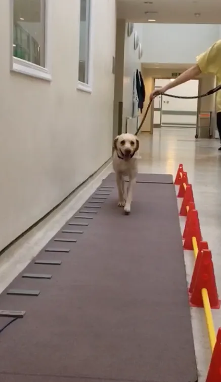
Evaluation of a lame patient can include a combination of assessment techniques, including subjective methods (eg, visual observation) and objective methods (eg, kinetics [measurement of force and pressure; Figure 1], kinematics [evaluation of movement of limb segments and joints]). In a clinical setting, visual observation is the most common evaluation technique because it is readily available and affordable and requires no additional equipment; however, visual gait analysis is subjective, has poor interobserver agreement (although it improves with lameness severity), and is poorly correlated with objective gait analysis.1,2 Kinetic and kinematic evaluation techniques are most frequently available in referral hospitals and research centers.

Dog walking on pressure-sensitive walkway for objective gait analysis
Gait analysis at a walk and a trot should be performed in all lame dogs. For patients with suspected CCLD, combining clinical measurements (eg, thigh circumference, joint range of motion, craniocaudal instability, joint effusion) and functional tests (eg, gait analysis, sit test, stair climbing) has been reported to have good correlation with objective kinetic evaluation at a trot.3 When objective assessment techniques are not available, a combination of clinical measures and functional tests can thus be helpful.
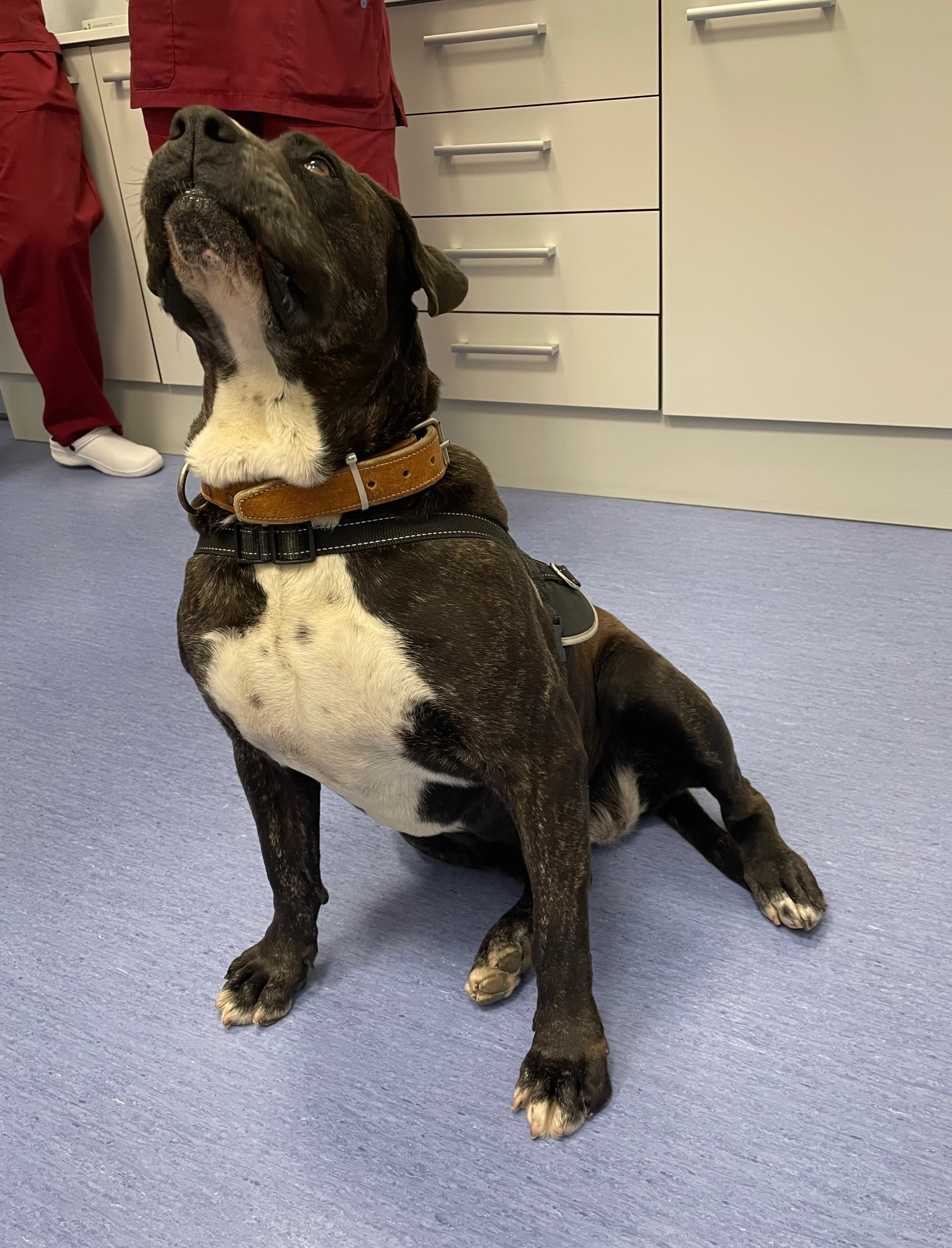
Patients with CCLD generally have instability, pain, joint effusion, and decreased range of motion in the affected stifle and frequently avoid full stifle flexion. Patients may sit with the affected limb abducted instead of sitting square (which requires full flexion of the stifle); this may be observed during a sit test (Figure 2). Although objective use of STST was not able to correctly identify all lame dogs with CCLD, subjective use along with other clinical and functional tests can be helpful.

Dog sitting with pelvic limb abducted during a sit test
You are reading 2-Minute Takeaways, a research summary resource presented by Clinician’s Brief. Clinician’s Brief does not conduct primary research.