Feline Pemphigus Foliaceus: A Common Autoimmune Dermatosis
Alexander Werner Resnick, VMD, DACVD, Animal Dermatology Center
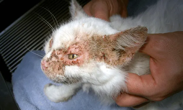
Classic pattern of feline pemphigus foliaceus with adherent crusts around the face, dorsal muzzle, periorbital region, and pinnae.
This companion article to “Canine Pemphigus Foliaceus,” which appeared in our June 2012 issue, presents an overview of this disease in cats. As in dogs, pemphigus foliaceus is the most common feline autoimmune dermatosis.
The primary lesions of pemphigus foliaceus are superficial pustules, which (as a result of being thin-roofed) frequently rupture to form crusts with underlying erosions. Coalescing regions of exfoliation and adherent crusts can become tender and exudative with development of secondary bacterial infection or when footpads are affected.
In cats, lesions most frequently develop at or around the nasal planum and muzzle, pinnae (Figure 1), nipples (Figure 2), ungual folds, and footpad margins (Figure 3). Exudates on the feet can be quite severe (Figure 4).

Thicklycrusted and erythematous plaque surrounding a nipple.

Excessive scaling of the footpads with crusts adherent to the footpad margins and nails.

Purulent-appearing exudate from the ungual fold.
The pathophysiology of feline pemphigus foliaceus involves development of autoantibodies, which produce a loss of intercellular adhesion in the upper layers of the stratum corneum (acantholysis). This leads to vesicle and pustule formation. Genetics, drugs (eg, antibiotics, methimazole), and other factors are known triggers for antikeratinocyte autoantibody induction.
Breed, sex, and age predilections have not been reported in cats.
Diagnosis

Cytologic preparation showing neutrophils and large rounded (acantholytic) keratinocytes.
Cytologic examination of pustule contents may provide a tentative diagnosis based on the presence of individual to rafts of immature and free-floating (acantholytic) keratinocytes, in addition to intact neutrophils and occasional eosinophils (See Figure 5, Cytologic preparation showing neutrophils and large rounded (acantholytic) keratinocytes). However, histopathologic examination of skin biopsy tissues (ie, intact pustule when possible) is required for definitive diagnosis.
Treatment
The treatment protocol requires immunosuppressive therapy. Although treatment for canine pemphigus foliaceus often requires multidrug protocols, administration of corticosteroids alone may be successful in cats. The author prefers treatment with dexamethasone in cats (0.22 mg/kg q24h at a tapering dose) as the initial corticosteroid choice. When combination therapy is instituted, chlorambucil (2 mg/m2 q48h) or cyclosporine (5 mg/kg q24h) may be added.
In general, secondary bacterial infections are a less significant concern in cats than they are in dogs. However, when bacteria are present in cytologic preparations, infections must be controlled with oral antibiotics and topical antiseptic bathing in the induction treatment phase. Cases complicated by previous antibiotic administration may require exudate culture and sensitivity testing for selection of appropriate medication.
Prognosis
Prognosis for feline pemphigus foliaceus is fair to good. As with dogs, affected cats benefit most from aggressive initial treatment that induces remission, followed by maintenance therapy for long-term control of the disease.