Canine Pemphigus Foliaceus
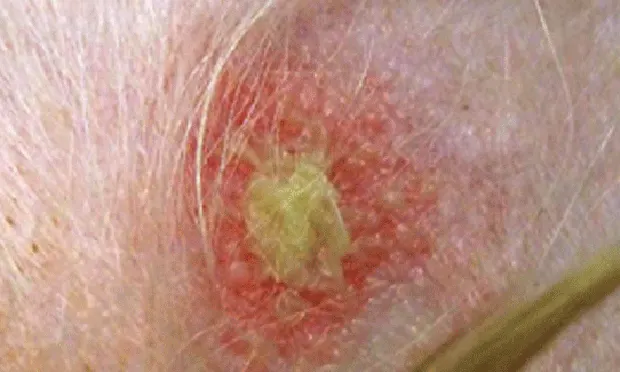
Pemphigus foliaceus, the most common autoimmune dermatosis in dogs, presents with primary large, superficial pustules. Pustules may be punctate or span multiple follicles, and contents may appear translucent to yellow (Figure 1). Because they are thin-roofed, pustules tend to rapidly rupture and form crusts with underlying erosions.
Figure 1 (above). Flaccid pustule of pemphigus foliaceus spanning multiple follicles.
Preceding the eruption of lesions, which often develop in waves, some patients experience a period of lethargy and inappetence. When a secondary bacterial infection develops or the lesions affect the footpads, coalescing regions of exfoliation and adherent crusts can become tender and exudative.

Several disease patterns are described, including a facial (classic butterfly) pattern affecting the nasal planum, dorsal muzzle, and periorbital and pinnal areas, as well as on the footpad margins and trunk (Figures 2 and 3).
Figure 2. Classic lesions of pemphigus foliaceus with thick crusts on the rostral muzzle, nasal planum, dorsal muzzle, and periorbital regions (butterfly pattern).

Antikeratinocyte antibodies, which have been identified in cases of canine pemphigus foliaceus, can cause loss of intercellular adhesion in the upper layers of the stratum corneum (acantholysis), leading to vesicle and pustule formation. Factors considered to be triggers for antikeratinocyte antibody induction include genetics and drugs (ie, antibiotics, phenobarbital, metaflumizone).
Figure 3. (A) Yellow adherent crusts on the pinna of a Maltese dog with pemphigus foliaceus. Lesions developed on both surfaces of the pinnae, including glabrous areas. (B) Pinna of same patient after 4 weeks of therapy. Lesions that were originally large accumulations of crusts are now punctate crusted papules and pustules.
DiagnosisTentative diagnosis is possible by examining pustule contents. Pustule cytology should reveal individual to multiple immature and free-floating (acantholytic) keratinocytes as well as intact neutrophils and occasional eosinophils. Definitive diagnosis requires histopathologic examination of skin biopsy tissue. An intact pustule is preferable.
TreatmentTreatment requires immunosuppressive therapy. Both single modality (ie, corticosteroid therapy) and multidrug treatment plans have been described, although concurrent use of corticosteroids with corticosteroid-sparing medications (eg, azathioprine) is recommended.
Especially during the induction phase of treatment, secondary bacterial infections must be controlled through oral antibiotics and topical antiseptic bathing. In cases complicated by prior antibiotic administration, exudate culture and sensitivity testing may be required to select the appropriate medication.
In severe cases, bathing daily with a sulfur/salicylic acid shampoo to gently soak off crusts and exudates can significantly improve patient comfort. This also allows better assessment of the response to therapy. Once the disease is controlled, bathing can be reduced to once or twice weekly.
PrognosisThe prognosis for canine pemphigus foliaceus is fair to good. Cases benefit most after initial aggressive treatment to induce remission, followed by reduced therapy to maintain control of the disease (Figure 3).
ALEXANDER WERNER, VMD, DACVD, is owner of Animal Dermatology Center in Studio City, California, with offices in Westlake Village, California, and Reno, Nevada. In addition to clinical practice, he has presented lectures and published numerous articles and book chapters on dermatology. He is coeditor of the latest edition of Blackwell’s Five-Minute Veterinary Consult: Small Animal Dermatology. Dr.Werner received a VMD from University of Pennsylvania and completed a dermatology residency at University of California, Davis.