In the Literature
Acevedo A, Pablo LS, Vettorato E. Complete mechanical expiratory obstruction due to expiratory valve malposition in an anesthetized dog. Vet Anaesth Analg. 2025;52(4):498-502. doi:10.1016/j.vaa.2025.04.008
The Research …
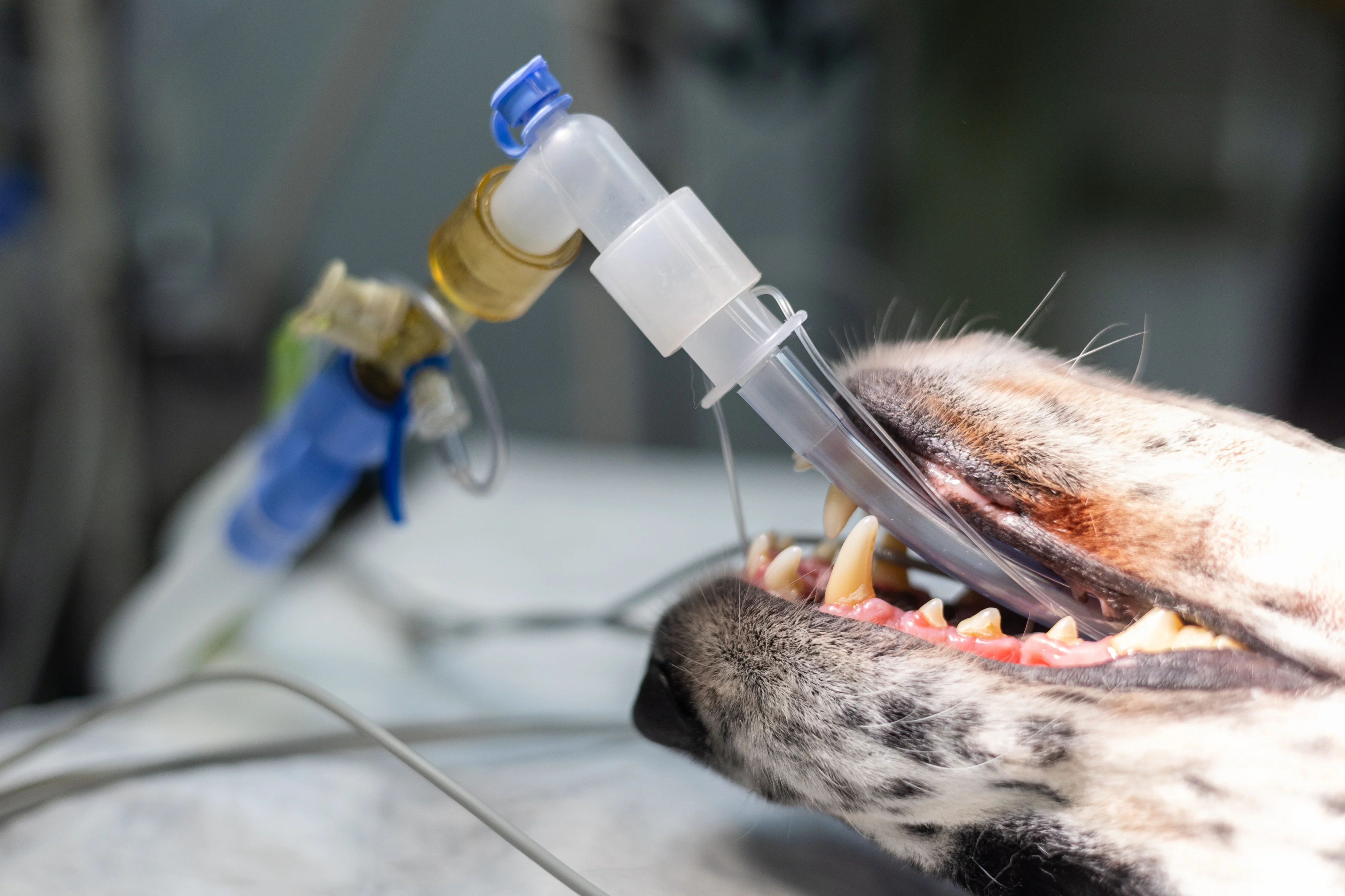
In this case study, an 8-year-old female crossbreed dog was presented for a tracheotomy and caudal segmental mandibulectomy. The anesthetic machine was set up with a rebreathing circuit and successfully leak tested with a closed system pressure of 20 cmH2O prior to use. Premedication and induction were uneventful. Tracheotomy was performed, followed by tracheal intubation with a cuffed endotracheal tube at the tracheotomy site.
When the endotracheal tube was connected to the breathing system, 2 manual breaths were given; however, no capnography waveform was seen despite attachment. The breathing system was disconnected for troubleshooting, and the anesthetist exhaled directly into the capnograph sampling adaptor, producing a normal capnograph waveform. The endotracheal tube was visualized to ensure there were no kinks.
When the capnograph adaptor was attached to the patient without the breathing circuit, the patient’s spontaneous ventilation displayed a normal capnograph waveform. The breathing circuit was reattached, and 2 additional manual breaths were given while the lung field was auscultated, confirming airflow; however, no capnograph waveform was seen. The breathing system manometer then increased rapidly to 20 cmH2O despite an open adjustable pressure limiting valve (often called the pop-off valve). The patient was transferred to a second anesthetic machine, which showed a normal capnograph waveform and a manometer reading of 0 cmH2O. The patient was maintained on this machine uneventfully for the remainder of the procedure, and her cardiovascular status remained within normal limits throughout the 7-minute troubleshooting period.
Following the procedure, the first anesthetic machine was examined closely, and the expiratory valve was observed to not move due to incorrect placement. The expiratory dome prongs were pressing the flutter valve against the expiration seat, which did not allow the valve to move during expiration. When this was corrected, the valve operated correctly. Prior assessment of unidirectional valve function can alert the team to this error; however, a standard machine leak test does not assess for this.
… The Takeaways
Key pearls to put into practice:
A standard preanesthetic system leak test can ensure there is no leak in the breathing circuit and machine; however, this test does not assess unidirectional valve function, which is performed via visual inspection of the valve. Blowing into the expiratory port should raise the expiratory unidirectional valve in a functional system.
Best practice to confirm endotracheal tube placement is via capnography. Other methods include direct visualization, placing a hand at the end of the endotracheal tube to feel for the presence of air while an assistant squeezes the patient’s chest, and connecting the patient to the anesthetic machine and auscultating the lung fields while an assistant delivers manual breaths.
The most common cause of expiratory obstruction is a kinked or obstructed endotracheal tube; however, an obstructed expiratory limb of the breathing circuit, malfunctioning expiratory valve, or occluded active scavenging system can also lead to dysfunction.1
Preanesthetic leak tests and monitoring equipment allow the anesthetist to provide a safe experience while minimizing anesthetic morbidity and mortality. The anesthetist should realize abnormalities quickly and troubleshoot in real time if complications arise.
You are reading 2-Minute Takeaways, a research summary resource presented by Clinician’s Brief. Clinician’s Brief does not conduct primary research.