Scaling & Crusting Skin Disease
Douglas J. DeBoer, DVM, DACVD, University of Wisconsin–Madison
Updated May 2025 by Charlie Pye, DVM, DVSc, DACVD; University of Prince Edward Island, Charlottetown, Prince Edward Island, Canada
The skin commonly develops scaling and crusting lesions in response to a variety of inflammatory or metabolic insults. Some characteristics of the lesions can be suggestive of a specific condition; in other situations, the appearance of the lesions may be nonspecific but clues can be derived from location, distribution, age of onset, breed, presence and degree of pruritus, and any other clinical signs.
Quiz: Scaling & Crusting Skin Disease
In the quiz below, match each disease with only one image and description. Some of the images may seem to fit well with more than one disease, but each image has only one correct match.
Scaling & Crusting Skin Lesions: Answers & Explanations
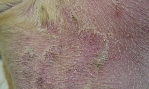
FIGURE 1 Diffuse, delicate scaling on the dorsum of a 2-year-old male golden retriever
Congenital Ichthyosis
Congenital ichthyosis is an uncommon hereditary and primary cornification defect in golden retrievers.1 A mutation in the PNPLA1 gene, thought to play a role in lipid organization and metabolism in the epidermis, has been documented.2 This image shows characteristic large and delicate rice paper scales on the trunk that generally occur without inflammation or pruritus and begin to appear at or near birth.