Ask the Expert: What is the recommended diagnostic and management approach for a patient presented with epistaxis?
Epistaxis in dogs is common, but published data remain limited. In a retrospective study of 176 dogs, reported local causes included nasal neoplasia (30%), trauma (29%), idiopathic rhinitis (17%), and periapical abscess (2%).1 Systemic causes included severe thrombocytopenia (10%), thrombocytopathy (6%), abnormal secondary hemostasis (3%), systemic hypertension (2%), and vasculitis (1%).1 Local and systemic causes may overlap, but neither duration of signs nor laterality can reliably distinguish these categories.1
Clinical Signs
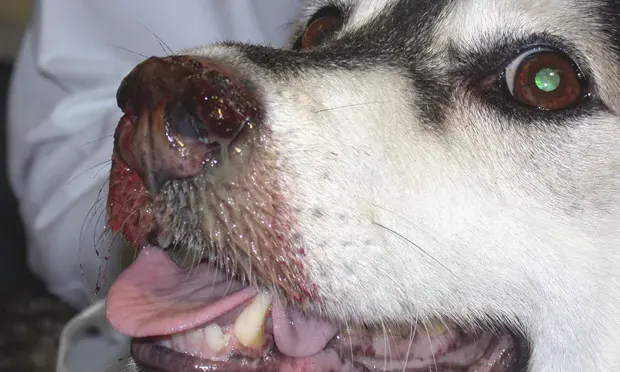
Epistaxis may be unilateral or bilateral and acute or chronic. Unilateral bleeding is more common and typically reflects the presence of local disease but is not pathognomonic for local disease. In one study, 52% of dogs with systemic hemostatic disorders exhibited unilateral signs.1 Additional signs may include nasal discharge, sneezing, stertor, altered nasal airflow, facial deformity, or systemic signs (eg, weakness, pallor).1 Behavioral changes (eg, altered mentation, aggressiveness) and seizures may occur with neoplasia or infectious/inflammatory disease (eg, encephalitis).2 Depigmentation and ulceration of the nasal planum may occur with fungal diseases, immune-mediated diseases, and neoplasia.
Causes
Epistaxis may result from local disease within the nasal cavity or systemic disease affecting hemostasis.
Local causes are predominant (present in up to 83% with dogs with epistaxis).1
Local Causes
Infectious
Fungal
Sinonasal aspergillosis is responsible for 12% to 34% of nasal disease in dogs.3 CT and rhinoscopy frequently reveal bilateral destructive rhinitis.3Patients with fungal rhinosinusitis (Aspergillus spp or Cryptococcus spp) can be presented with signs associated with sneezing, nasal discharge (including epistaxis), and stertor (cats).4,5
Parasitic
Cuterebra larvae and leech infestation have been reported as causes of chronic epistaxis.6,7
Angiostrongylosis
Patients with Angiostrongylus vasorum infection are at risk for bleeding even with unremarkable coagulation times. The underlying mechanisms are still poorly understood, and some patients may display severe hypercoagulable or hypocoagulable states secondary to disseminated intravascular coagulation, thrombocytopenia, hyperfibrinolysis, or other complex interactions. Respiratory signs (eg, cough, respiratory distress) are the most common reason for presentation.7,8
Leishmaniosis
In dogs with relevant travel history or potential exposure, Leishmania spp can cause epistaxis secondary to thrombocytopathy (ie, dysfunction of platelets), hyperviscosity from hyperglobulinemia, or rhinitis.9,10
Neoplastic
Tumors of the nasal cavity and paranasal sinuses account for 1% to 2% of all canine tumors.3,11 Carcinomas represent nearly two-thirds of canine intranasal tumors and are characterized by a progressive local invasion with low metastatic rate at diagnosis. Epistaxis is reported in up to 69% of these cases.12 Other common sinonasal tumors in older, large-breed dogs include osteosarcoma and chondrosarcoma.11 Rare tumors include leiomyoma, intravascular lymphoma, and transmissible venereal tumors.13-15
Nasal neoplasia is uncommon in cats, accounting for <10% of all tumors, with lymphoma diagnosed most frequently, followed by adenocarcinoma and squamous cell carcinoma. Risk increases with age, and neutered male cats may be overrepresented. Rare tumors include neuroendocrine neoplasms and olfactory neuroblastomas.3
Inflammatory Causes
Dental Disease
The oral cavity should be carefully evaluated for dental disease, including tooth root abscesses and oronasal fistulas.
Foreign Body
Acute sneezing is common after inhalation of foreign material. Grass awn rhinitis can mimic nonforeign-body rhinitis, with unilateral discharge being most common, but 14% of dogs develop bilateral discharge and 9% exhibit epistaxis.6
Idiopathic Lymphoplasmacytic Rhinitis
Idiopathic lymphoplasmacytic rhinitis is a common chronic nasal disease in dogs.16 Clinical signs may include epistaxis, mucoid or serous nasal discharge, reverse sneezing, stridor, and inspiratory dyspnea. Most patients are presented with unilateral or bilateral nasal discharge.16
Steroid-Responsive Meningitis Arteritis
Steroid-responsive meningitis arteritis is an immune-mediated inflammatory disease that affects the meninges and associated arteries and is characterized by pyrexia, lethargy, and acute cervical pain. Although epistaxis is not usually reported as a finding of steroid-responsive meningitis arteritis, systemic vasculitis involving vessels in the nasal area can cause epistaxis.17
Nonneoplastic Masses
Nasal hamartomas are rare, nonneoplastic masses composed of disorganized native tissue. Epistaxis is a common sign and occurs in up to 66% of reported cases.18
Trauma
Traumatic epistaxis is typically acute and accompanied by other signs of injury.1 Obtaining a complete patient history can help determine the cause.
Systemic Causes
Primary Hemostatic Disorder
The most common platelet disorder is thrombocytopenia, followed by von Willebrand disease (vWD).19 With thrombocytopenia, the number of platelets is directly related to the clinical risk for bleeding. Causes can include decreased production, increased consumption, sequestration, increased destruction, and loss. vWD is an inherited disease associated with structural and quantitative deficiencies in von Willebrand factor (vWF), which attaches platelets to injured vascular endothelium and allows platelets to aggregate; vWD can result in increased bleeding tendencies.20
Coagulation Factor Deficiency
Congenital causes include hemophilia A and B. Hemophilia A is a deficiency of factor VIII and the most commonly inherited deficiency of coagulation. Hemophilia B (more rare than hemophilia A) is a deficiency in factor IX and a recessive trait linked to the X chromosome.20 Acquired causes include exposure to anticoagulant rodenticide (which is not allowed in products intended for residential use), liver failure, and disseminated intravascular coagulation.
Paraneoplastic Syndrome
Paraneoplastic hyperfibrinolysis can occur when tumor cells produce profibrinolytic proteins (eg, urokinase-type plasminogen activator, tissue plasminogen activator). Hyperfibrinolysis is an uncommon but potentially fatal paraneoplastic manifestation that has been associated with prostate cancer, breast carcinoma, and sarcoma, as well as esophageal adenocarcinoma in humans.21,22 A case secondary to metastatic nasal adenocarcinoma has been reported in an 8-year-old neutered male border collie.7
Infectious
Canine systemic aspergillosis is a rare but life-threatening fungal disease characterized by hematogenous dissemination of Aspergillus spp and involves multiple organs and systems, including respiratory, GI, nervous, musculoskeletal, urinary, lymphatic, and ocular systems. German shepherd dogs are the most frequently affected breed.23
Diagnosis
A structured diagnostic approach is recommended. Initial evaluation should include packed cell volume, total protein, CBC with blood smear evaluation, serum chemistry profile, coagulation testing (including prothrombin time [PT] and activated partial thromboplastin time [aPTT]), and blood pressure measurement.24 In patients with severe epistaxis, assessment of the severity of perfusion impairment should include perfusion parameters (eg, heart rate, pulse quality, mucous membrane color, capillary refill time, peripheral temperature, mentation).
CBC
CBC may be normal in some patients with epistaxis, but the presence of regenerative anemia indicates bone marrow response to bleeding.
Leukocytosis is nonspecific, and leukopenia may also be present.
A blood smear evaluation can be useful to determine platelet count while laboratory results are pending and can indicate the presence of macroplatelets.
Automated platelet counts can be determined via a commercial laboratory analyzer; however, errors are common and can overestimate or underestimate the automated platelet count.
Manual platelet counts can be performed via evaluation of a peripheral blood smear using EDTA blood or fresh samples.
The sample should be evaluated on 100× high-powered field to assess the monolayer.
Each platelet is equivalent to 15 × 103/μL platelets. The platelet average in 5 fields can be multiplied by 15 × 103/μL to obtain the platelet estimation.25
If the platelet count is normal but signs are consistent with a primary hemostatic disorder, platelet function testing should be considered. Buccal mucosal bleeding time, platelet function analysis, vWF assay, and aggregometry tests can evaluate the ability of platelets to adhere, aggregate, and/or activate.19,26
Serum Chemistry Profile
Panhypoproteinemia can result from chronic blood loss, neoplasia, or infection.
Hyperlactatemia can be a sign of moderate to severe hemorrhagic shock.27
Blood Pressure Measurement
In cases in which hypertension is suspected to be a contributing factor of epistaxis, blood pressure should be evaluated using indirect Doppler or oscillometric methods.28
Coagulation Testing
Viscoelastic coagulation assays can provide a global assessment of clot formation kinetics and strength, in which platelet function plays a pivotal role. Tests for secondary hemostasis include PT and aPTT.
PT evaluates the extrinsic and common coagulation pathways and may be prolonged in patients with factor deficiency or dysfunction, including vitamin K-dependent coagulation factors altered because of anticoagulant rodenticide ingestion.
aPTT is a screening test that evaluates the intrinsic and common coagulation pathways and is not affected by primary hemostatic disorders.
Activated clotting time is a simple, rapid, inexpensive test that assesses for all clinically significant clotting factors, except factor VII. The activated clotting time may be mildly to moderately prolonged in patients with severe thrombocytopenia and some thrombopathies.8
Imaging & Airway Evaluation
Imaging should be performed prior to other diagnostics because of the potential for procedure-induced hemorrhage.
CT is the first-line modality for suspected neoplasia or destructive rhinitis. Although advanced imaging can provide significant information and increased diagnostic sensitivity in dogs with nasal disease compared with plain radiography, cost and availability can be limiting.
Radiography of the skull may have poor to moderate sensitivity in detection of soft-tissue changes and moderate sensitivity in detection of bony changes and sinus evaluation.
Rhinoscopy allows direct visualization of the nasal passages and choanae and can help identify parasites, foreign material, fungal plaques, stenosis/atresia, and masses.29
Upper airway examination should be performed with the patient under general anesthesia and is often combined with rhinoscopy. Saline flushing of the nasal cavity may be considered for diagnostic or therapeutic purposes.30
Thoracic imaging can be considered in dogs with suspected nasal neoplasia or fungal disease for visualization of a metastatic process or systemic involvement.28,29
Additional Diagnostics
Additional diagnostics include PCR and/or serology for infectious causes, cytology, and histopathology. Infrared thermography is a novel, noninvasive, portable tool used to detect surface temperature patterns associated with nasal obstruction.31
Treatment
Immediate Stabilization & Local Therapies
Triage examination should focus on perfusion parameters and systemic stability. Initial approaches may include nasal packing with sponges, sedation to reduce anxiety and blood pressure, cold compresses, and treatment of the underlying cause.27 Specific treatment for severe epistaxis may be attempted, particularly when initial approaches and topical vasoconstrictors (eg, epinephrine, phenylephrine) are unable to control hemorrhage.9
Systemic Therapies
Fresh whole blood is indicated in patients with hemorrhage caused by severe thrombocytopenia, platelet disorders (congenital or acquired), and vWD with anemia, whereas cryoprecipitate may be indicated in patients with vWD without anemia.32 Lyophilized platelets can be considered for prevention of bleeding in dogs with thrombopathy or thrombocytopenia.33
Antifibrinolytic drugs (eg, aminocaproic acid, tranexamic acid) can be applied locally or systemically, are indicated for thrombocytopenia with associated hemorrhage (eg, epistaxis), and can be used in combination with platelet transfusions. IV aminocaproic acid can be administered at 50-100 mg/kg every 6 hours or 50 mg/kg bolus followed by an infusion of 15 mg/kg/hour over 8 hours. Oral aminocaproic acid can be administered at 100 mg/kg every 6 hours or 500-1,000 mg every 8 hours after 15-40 mg/kg IV has been given. Tranexamic acid can be administered at 15-20 mg/kg PO every 8 hours, at 20 mg/kg IV every 6 to 8 hours, or as a 20 mg/kg IV bolus followed by an IV infusion of 20 mg/kg over 8 hours.27,34
Desmopressin can enhance platelet aggregation, supplement vWF deficiencies, increase endothelial vWF release to improve platelet adhesion, and shorten bleeding times through vWF‑independent mechanisms.12 Desmopressin is indicated in patients with hemophilia A and vWD (thrombocytopathies) and can be used to stop spontaneous hemorrhage. Desmopressin may be administered at 1-5 micrograms/kg SC using the intranasal formulation or at 0.3-1 micrograms/kg IV or SC using the parenteral product.27
Additional therapy depends on the underlying condition and may include whole blood, packed RBCs, fresh frozen plasma, cryoprecipitate, or vitamin K.27,35
Advanced or Refractory Options
Permanent ligation of the common carotid artery may provide temporary benefit due to collateral revascularization. Percutaneous arterial embolization can be performed by positioning a microcatheter just proximal to the origins of the minor palatine artery, the common trunk of the sphenopalatine and major palatine arteries, and the infraorbital artery.36,37
An improvised nasopharyngeal tamponade device (ie, condom affixed to a nasogastric tube) was tested in cadavers to compress bleeding vessels and allow intranasal drug instillation of hemostatic medications (50% diluted ioversol) without leakage and may be considered as a low resource alternative; however, this has not been tested in actively hemorrhaging patients.38
In patients with nasal bleeding after endoscopic surgery, different alternatives can be considered for nasal packing material. In a model of canine nasal bleeding, high-expansion degradable cotton was trialed as a degradable nasal hemostatic material after endoscopic surgery as an alternative to other materials.39
Balloon catheter placement within the nasal passage has been described to provide local pressure and has been used in combination with carotid artery ligation in a case of severe epistaxis secondary to leishmaniosis.13
Definitive Treatment
Definitive treatment focusing on management of the underlying cause (eg, antifungal therapy for aspergillosis, chemotherapy or radiation for neoplasia, immunosuppression for immune-mediated thrombocytopenia) should be pursued.