Diagnostic Challenges of Feline Heartworm Disease
Ashley B. Saunders, DVM, Diplomate ACVIM (Cardiology), Texas A&M University
Heartworm disease has emerged as an important feline disease that is challenging to both diagnose and treat. Cats are not the typical host for Dirofilaria immitis and are inherently resistant to infection, yet the importance of recognizing the disease in cats cannot be overemphasized.
Related Article: Coughing Cat: Could It Be Heartworm?
On average, cats have a small heartworm burden (typically 1 to 3 worms) and are rarely microfilaremic. Clinical signs of feline heartworm disease are often nonspecific (see below). Cats are at risk for thromboembolism and aberrant heartworm migration to the eyes and brain, resulting in blindness and seizures.
Recent research has documented pulmonary changes contributing to clinical signs in the early stages of the disease. The clinical syndrome, referred to as heartworm-associated respiratory disease (HARD), is characterized by pulmonary lesions and clinical signs associated with larval and adult heartworms that are often diagnosed as feline asthma. The nonspecific clinical signs and potential for pathologic events early in the course of disease before a positive test result can be obtained highlight the importance of administering heartworm prevention to cats.
Historical Findings in Cats with Heartworm Disease
Cough
Lethargy
Dyspnea
Vomiting
Anorexia
Blindness
Weight loss
Seizures
Levels of Care
Cats that live outdoors are at increased risk for heartworm disease; however, it is important to remember that cats living exclusively indoors are also at risk. The nonspecific nature of the clinical signs associated with heartworm disease makes diagnosis challenging, and an index of suspicion for heartworm disease is often the first step in diagnosis.
Diagnosing heartworm disease in cats frequently requires a combination of tests. Serologic testing can be useful for initial screening purposes but is often inadequate. A positive heartworm antibody test is significant, as it indicates previous or current infection with heartworms and a potential increased risk for the disease. Since heartworm antigen tests are only positive in the presence of female worms, they may yield false-negative results. In addition, different antibody tests identify different heartworm proteins expressed at different life stages; false-negative results may be elicited in the presence of larval or adult stage heartworms.
Related Article: Heartworm Infection in Cats
Supportive evidence of heartworm disease may be present on additional diagnostic tests. Although cats are rarely microfilaremic, microfilaria may be detected on a blood smear. Eosinophilia is often transient and rarely identified. Thoracic radiographic findings in cats with heartworm disease include enlarged pulmonary arteries, especially the right caudal lobar artery; bronchointerstitial infiltrate and occasionally pulmonary overinflation, pleural effusion, and pneumothorax may also be present. Radiographic abnormalities may be transient, and right heart enlargement is rarely identified.
Echocardiography can be a sensitive means for detecting heartworms but often requires a skilled ultrasonographer. Adult heartworms in cats are smaller than those found in dogs and typically reside in the pulmonary vasculature where imaging is difficult. Heartworms resemble hyperechoic parallel lines and may be identified in the pulmonary arteries, right ventricle, across the tricuspid valve, or within the right atrium.
When to Consider Referring
Referral may be helpful when heartworm disease is suspected but the diagnosis has not been readily established. Referral may also be considered for echocardiographic evaluation by an ultrasonographer adept at imaging cats, especially to assess cats with murmurs, arrhythmias, or potential caval syndrome.
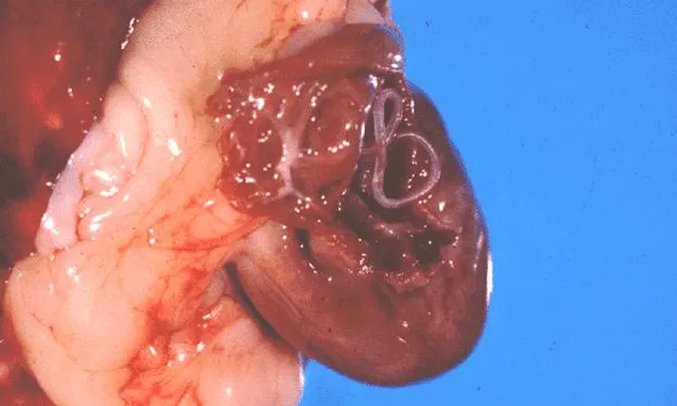
Echocardiographic evidence of heartworms within the right atrium or across the tricuspid valve, as occurs with caval syndrome, in combination with clinical signs of heartworm disease should prompt referral to a facility capable of heartworm extraction. Caval syndrome is uncommon in cats; surgical extraction has been successfully used to treat this condition but is not risk-free.
The Referral Process
Cats with respiratory distress should be stabilized before referral to prevent decompensation or death during transport. Recent blood analysis results, radiographs, and any other pertinent medical records should be sent with the patient. Owners should be prepared that cats with respiratory distress are at high risk for decompensation and sudden death. Many cats can be managed medically during the life of the heartworms (2 to 4 years) and maintain a good quality of life during that time.
When Referral Is Not an Option
Treatment for cats with persistent dyspnea or episodic respiratory distress requires facilities with 24-hour care; however, cats should not be transported until stabilized, which is the primary goal of management.
Cats with respiratory distress often respond to medical management using oxygen supplementation, corticosteroids (dexamethasone, 1 mg/kg IV or IM, or tapering doses of prednisone, 1-2 mg/kg PO Q 12-24 H), and bronchodilators (terbutaline, 0.01 mg/kg SC Q 4 H; aminophylline, 6.6 mg/kg PO Q 12 H; or sustained-release theophylline tablets, 15 mg/kg PO Q 24 H). It may be prudent to provide owners of cats with known heartworm disease a supply of steroids in case an acute crisis occurs at home.
Doxycycline has been suggested as possible concurrent therapy for heartworm-infected cats to treat Wolbachia, a bacteria associated with heartworm infections that is considered to be the trigger for acute reactions in dogs and cats. However, evidence of improved outcome with concurrent use of doxycycline in treating cats with heartworm infection is inconclusive at this time. Evaluation in a clinical setting is required to fully elucidate the role of Wolbachia and doxycycline in this disease.
Case Study: Feline Heartworm Disease
A 2-year-old spayed female domestic shorthair cat was referred for intermittent respiratory distress and vomiting. The cat was housed indoors exclusively.
Upon examination, she had open-mouth breathing and a respiratory rate of 80 breaths/minute. Bronchovesicular sounds were harsh bilaterally without crackles or wheezes. Her mucous membranes were pink, and capillary refill time was within normal limits. She did not have a murmur or arrhythmia.
Right lateral (A) and dorsoventral (B) thoracic radiographs were taken. Notice the enlarged pulmonary artery (C, arrow) in the dorsoventral view, which provided a clue to her diagnosis.
Echocardiography was performed (D); the image is a left parasternal view of the right atrium (RA ) and right ventricle (RV ). Note the hyperechoic parallel lines representing heartworms in the RA and RV.
Results of heartworm antibody and antigen tests were both positive.


