Congenital Hepatobiliary Diseases in Dogs & Cats
Profile
Definition
Congenital hepatobiliary disorders include1-9
Vascular disorders (eg, congenital portosystemic shunt [CPSS], primary hypoplasia of the portal vein [PHPV], portal vein atresia, hepatoarteriovenous malformation [HAVM])
Biliary disorders (eg, ductal plate abnormality [DPA])
Inborn errors of copper metabolism
CPSS is a macroscopic connection between portal and systemic circulation.
Most common congenital hepatobiliary disorder in dogs
Can be intra- or extrahepatic
PHPV is a microscopic intrahepatic vascular abnormality in which portal venous blood is diverted into the hepatic veins within the intrahepatic microcirculation.
Formerly microvascular dysplasia (MVD); may occur with portal hypertension
PHPV without portal hypertension common in dogs
PHPV with portal hypertension (ie, noncirrhotic portal hypertension) rare
HAVM is failure in differentiation of embryonic venous and arterial structures (rare).
Portal vein atresia is the congenital absence or atresia of the extrahepatic portal vein (rare).
Copper-associated hepatic disease is an inborn error of metabolism in which copper transport in the liver is impaired or overwhelmed.
Common in dogs, but rare in cats
A DPA (ie, fibropolycystic disease) is an anomalous bile duct development at various levels of the biliary tree leading to persistent, aberrant ducts, hepatic fibrosis, and/or cyst formation.
Rare in dogs and cats, but autosomal recessive polycystic kidney disease (ARPKD) may occur in Persian cats
Bilobed gallbladder has been reported in cats and gallbladder agenesis in dogs.2,10,11
Both are incidental findings.
Gallbladder agenesis is possibly associated with DPA.
Related Article: Top 5 Liver Conditions in Dogs
Signalment
Seen worldwide in dogs and cats, but vascular disorders and copper-associated disease more common in dogs
PHPV has not been reported in cats.
Breed Predilection
CPSS
Intrahepatic is seen in large-breed dogs, Irish wolfhound (genetic, polygenic, patent ductus venosus), Australian cattle dog (usually right divisional), and Labrador retriever.3,12
Age of onset of signs typically <1 year
Extrahepatic is seen in Maltese, silky terrier, Yorkshire terrier, cairn terrier (autosomal, polygenic with variable expression), shih tzu, bichon frise, miniature schnauzer, pug, Havanese, and dandie dinmont terrier.3,12
Age of onset of signs typically 2 months–4 years (88% of cases); may be older
Incidence of 0.18% in purebreed and 0.05% in crossbreed dogs3
Most common congenital liver disease and more frequent in dogs
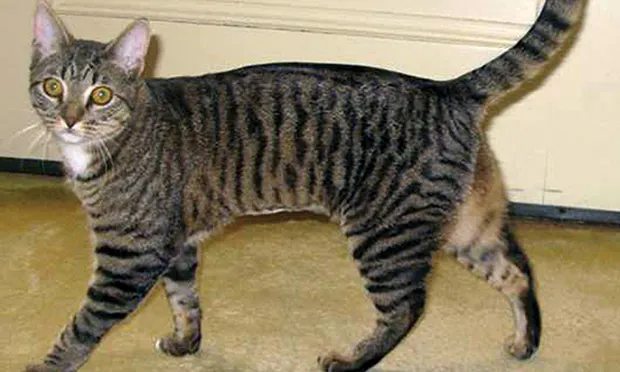
Affected cats often have a copper color to eyes (Figure 1).
Figure 1. Castrated domestic short-haired cat (10 months of age) 1 month after attenuation of portocaval shunt via placement of ameroid constrictor. The cat had uneventful surgery and was clinically normal 2 years later. Note the copper color to the eyes.
PHPV
Affects cairn terrier, Tibetan spaniel, Maltese, Havanese, Yorkshire terrier, Norfolk terrier, and miniature schnauzer
Allelic trait and may be an ancestral founder mutation
Age of onset of signs is typically <1 year but may go undiagnosed (many dogs asymptomatic)
In concurrence with portal hypertension, has increased incidence in Doberman pinscher, Cocker spaniel, and rottweiler; median age of onset of signs is 1–2 years6,13,14
Related Article: Interpretation of Serum Transaminase Levels in Dogs & Cats
HAVM
Age of onset of signs typically <1 year1
Copper-Associated Hepatic Disease
Bedlington terrier is usually diagnosed by hepatic copper quantification testing at 1 year of age; younger dogs may be diagnosed by genetic testing.
Affects middle-aged or older Dalmatian, West Highland white terrier, Skye terrier, Doberman pinscher, and Labrador retriever
DPA
Age at diagnosis depends on severity of lesion15-20
65% liver involvement in cats with mutation in polycystin gene
38% incidence for autosomal dominant polycystic kidney disease (ADPKD) in Persian and Persian-crossbreed cats21
ADPKD rare in West Highland white and cairn terrier19,22
Pathophysiology
Dogs with CPSS have shunting of portal blood into systemic circulation and hepatic atrophy from loss of normal hepatotropic factors in portal blood.
Portosystemic shunting of portal blood can lead to increased circulating levels of toxins originating from the intestinal tract (eg, hyperammonemia).
Ammonia is toxic to neuronal tissues and can cause hepatic encephalopathy.
Urolithiasis also occurs in animals with CPSS caused by ammonium biurate stones secondary to chronic hyperammonemia.
Dogs with PHPV have microscopic shunting of blood that typically does not cause liver damage.
PHPV rarely seen as more progressive disorder associated with vascular hypoperfusion that can cause resistance to portal blood flow and portal hypertension
Dogs may be born with aplastic extrahepatic portal vein, typically leading to portal hypertension.
Dogs and cats with HAVM typically have portal hypertension because of increased blood flow; concurrent PHPV common in dogs

Pathophysiology of DPA in dogs and cats is not well understood
Reports of congenital hepatic fibrosis have indicated that DPA can progress to portal hypertension and hepatic failure.
In dogs, may be similar to Caroli disease in humans.
Cats with ADPKD have varying cystic hepatic involvement but are more likely to present with progressive renal failure.
Animals with choledochal cysts (Figure 2) often present with secondary bacterial infections.16
In Bedlington terrier, hepatic copper accumulation related to autosomal recessive mutation in the COMMD1 gene (codes for a protein involved in intracellular copper transport)
Nature of genetic defect in other breeds not identified
Increased hepatic copper stores in Labrador retriever may be related to increased copper availability in dog foods.
If not treated, excess accumulation of copper in the liver causes oxidative damage to hepatocytes, which eventually results in cirrhosis and portal hypertension.
Portal hypertension is caused by increased resistance, increased blood flow, or both into normally low resistance portal circulation.
Mostly caused by hepatic parenchymal fibrosis
Clinical consequences include multiple acquired portosystemic shunts (MAPSSs), ascites, and/or hepatic encephalopathy.
Can occur with vascular disorders (eg, portal vein atresia, PHPV, HAVM), DPA, and copper-associated hepatic disease
Dogs with CPSS do not have portal hypertension.
Figure 2. Ultrasonography showing choledochal cysts in a domestic short-haired cat (5 years of age, A). The cat’s common bile duct (CBD) was greatly enlarged (cursors) and half filled with echogenic sediment. On previous ultrasounds, the CBD had been mistaken for the gallbladder and a hepatic abscess. The cat had recurrent bacterial cholecystitis until the malformation was surgically corrected. In a 10-month-old dog (B), the CBD is distended with echogenic walls. This dog also had gallbladder agenesis and congenital hepatic fibrosis. Bile culture was negative.
Clinical Signs
CPSS
In dogs with hepatic encephalopathy, episodic neurologic signs consistent with diffuse cerebral disease may vary in severity (eg, mild depression and behavioral changes to stupor, coma, seizures, blindness).
Ptyalism is seen in cats.
PU/PD, stranguria/pollakiuria, chronic intermittent vomiting and/or diarrhea, and poor BCS also possible
PHPV
Animals will be asymptomatic.
PHPV with Portal Hypertension
Vomiting, diarrhea, icterus, PU/PD, depression, lethargy, ascites, and hepatic encephalopathy
HAVM
Portal hypertension with ascites and hepatic encephalopathy
DPA
Chronic vomiting, diarrhea, PU/PD, hepatic encephalopathy, ascites with congenital hepatic fibrosis, or Caroli disease
In DPA confined to extrahepatic bile ducts (eg, choledochal cysts), signs of cholecystitis (ie, acute onset vomiting, icterus, fever, pain of the right abdominal quadrant) possible
Diagnosis
Definitive
In patients with CPSS, HAVM, and portal vein atresia, diagnostic imaging or surgical confirmation of vascular abnormality is needed.
DPA requires imaging of the abnormality and liver biopsy.
PHPV is a diagnosis of exclusion.
The only abnormality may be mild-to-moderate increases in pre- and/or postprandial total serum bile acid concentrations.
With portal hypertension requires combination of laboratory studies, imaging results, and histopathologic findings
Copper-associated hepatic disease requires examination of liver biopsy specimen and measurement of hepatic copper concentrations
Quantitative measurement >1500 ppm; qualitative measurement shows centrilobular distribution.
Differential
Infectious, toxic, inflammatory, or obstructive disease (See table: Differential Diagnosis of Canine Congenital Hepatic Disease)
Imaging
Radiography may show microhepatica, renomegaly (CPSS, DPA), cystoliths, calcification of biliary tract (DPA), and abdominal effusion with portal hypertension.23
Abdominal ultrasonography
In the liver, microhepatica with poor vascular marking (CPSS, HAVM) and shunts or HAVM shown
Microhepatica with nodular appearance and rounded edges presents in late-stage copper-associated liver disease
With extrahepatic CPSS, turbulence in caudal vena cava and portal:aortic ratio of <0.8 present24
Portal hypertension can lead to abdominal effusion, decreased flow velocity in portal vein, hepatofugal flow in portal vein, and MAPSS.
Renomegaly and kidney or bladder stones associated with CPSS; cysts associated with DPA
With DPA, cystic dilation of intrahepatic or extrahepatic biliary system shown
Signs of biliary infection are possible: biliary sludge, cholelithiasis, thickened or calcified bile ducts.
Inability to visualize gallbladder may result from gallbladder agenesis.
Copper-associated hepatic disease causes hepatomegaly that progresses to microhepatica with nodule formation and irregular edges.
Transcolonic or transplenic scintigraphy normal with PHPV if portal hypertension absent23
Increased shunt fraction possible with CPSS or MAPSS
Differentiation between congenital or acquired shunting may be impossible.
CT or MRI angiography provide greater vascular detail; quickly replacing intraoperative portovenography as gold standard for visualization of portal vasculature23
Laboratory Findings
CBC results and examination of blood smear show microcytosis (CPSS, MAPSS) and normochromic, nonregenerative anemia.
Leukocytosis uncommon but associated with poor prognosis (CPSS) or complicating cholecystitis (DPA)
Serum chemistry panel shows mild-to-moderate elevations in ALP, ALT, AST and decreased blood urea nitrogen, albumin, and cholesterol concentrations.
Hypoglycemia possible in toy breeds with CPSS
Increased total bilirubin concentration associated with PHPV with portal hypertension, DPA, and copper-associated hepatic disease, but rare with CPSS
Urinalysis shows ammonium biurate crystalluria; isosthenuria or hyposthenuria with PU/PD; and hematuria, pyuria, or bacteriuria with infections secondary to stones.
Measurement of pre- and postprandial serum bile acid concentrations can screen for vascular disease.
Postprandial measurement most sensitive (concentrations should be >25 µmol/L)
Increased values with DBA and copper-associated hepatic disease in late stages
Increased blood ammonia concentrations in diseases associated with vascular shunting
Protein C values within range with PHPV and decreased with CPSS or acute hepatic failure (rare manifestation of copper-associated hepatic disease)
Abdominal fluid analysis shows decreased protein concentration (<2.5 g/dL) and pure or modified transudate.
Hepatic biopsy
Chronic hypoperfusion possible to varying extent with CPSS, PHPV, HAVM, portal vein atresia, and DPA
Typical histopathologic features of chronic hypoperfusion include underdeveloped portal triads, hepatic arteriolar reduplication, lobular atrophy, dilation of perivenular lymphatics, bile duct proliferation, and multifocal lipogranulomas.
Centrilobular fibrosis rare with CPSS or PHPV
Accurate histopathologic diagnosis often requires wedge biopsy specimens from multiple liver lobes, as microscopic lesions can be patchy and subtle.
With PHPV with portal hypertension, varying degrees of periportal and/or centrilobular fibrosis possible, but without inflammation or regenerative nodules
With DPA, congenital hepatic fibrosis characterized by diffuse periportal to bridging fibrosis with numerous small irregular immature-appearing bile ducts
Mild arteriolar proliferation and portal vein attenuation
With Caroli disease, ADPKD (or ARPKD), multiple unilocular or multilocular biliary cysts present.
Centrilobular hepatocyte degeneration and lipidosis with copper accumulation (rhodanine staining) in early copper-associated hepatic disease
In later disease, centrilobular hepatitis progresses to periportal areas with associated inflammation and fibrosis; this eventually progresses to cirrhosis with regenerative nodules.
Treatment
Treatment based on whether underlying anomaly is amenable to interventional correction (eg, CPSS, HAVM, some choledochal cysts).
Unknown whether CPSS have same success by medical surgical means25
Medical Management
Goals targeted at controlling signs
With hepatic encephalopathy, treatment targeted to increase protein tolerance with lactulose and/or antibiotics (eg, metronidazole, neomycin)
Protein intake should be optimized by providing adequate protein and calories to avoid muscle catabolism.
High-quality protein derived from plant, dairy, or soy products preferred; fiber content should be increased.
Precipitating factors (GI bleeding, metabolic alkalosis, overuse of sedatives and analgesics, concurrent infections, azotemia, constipation) should be controlled.
For ascites from portal hypertension, restriction of dietary sodium and initiation of diuretics (ie, spironolactone, +/- low-dose furosemide) indicated
GI ulceration or bleeding should be addressed with H2-receptor blockers or proton-pump inhibitors.
Copper-associated hepatic disease requires copper chelation therapy (ie, penicillamine, trientine), decreased copper absorption (ie, administration of zinc), and low-copper diet.
Hepatoprotectants (eg, vitamin E, S-adenosylmethionine, ursodeoxycholate) recommended
For DPA, manage infectious complications and administer hepatoprotectants.
PHPV does not require treatment if portal hypertension not present; hepatoprotectants may be administered if liver enzyme values abnormal.
Surgery
For extrahepatic CPSS, attenuation by ligation or placement of ameroid constrictor or cellophane band indicated3,25
In dogs, perioperative mortality can be ≤10%; significant morbidity (eg, portal hypertension, postligation neurologic syndrome, thrombosis) seen
Levetiracetam pretreatment may decrease neurologic complications.13
Long-term outcome difficult to assess; studies have used diverse outcome measures with limited follow-up.
With complete ligation, ameroid constrictor, or band placement, recurrence of signs seen in ~10% of patients
Recurrence higher for partial ligation (40%–50%)
Cats have greater perioperative mortality (≤20%) and morbidity (neurologic complications in ≤37% of patients).
Only about 45% of cats have excellent or good outcome.
Surgical morbidity and mortality are higher with intrahepatic CPSS and thus interventional radiology is recommended.26
Interventional Radiology
Fluoroscopic-assisted percutaneous transvenous coil embolization is performed for intrahepatic CPSS; this procedure is considered as effective as and safer than surgical attenuation.27
Cyanoacrylate glue occlusion of HAVM is associated with a better outcome than surgery.
For choledochal cyst, monitor extirpation or marsupialization into the common bile duct; this improves bile flow and prevents recurrent cholangitis.
Follow-up
Monitoring for resolution of signs after withdrawing medications indicated
Normalization of pre- and postprandial total serum bile acid concentrations may not occur with concurrent PHPV.
Decreased shunt fraction seen on scintigraphy
Ultrasound verification of shunt closure indicated
In General
Relative Cost
Without surgical intervention: $$$–$$$$
With surgical intervention: $$$$$
Prognosis
CPSS, HAVM: varies
DPA: unknown—prognosis may depend on age at first appearance of clinical signs.
PHPV without portal hypertension: excellent
PHPV with portal hypertension: some dogs may “outgrow” disease and have prolonged survival.
Copper-associated hepatic disease: good if diagnosed early
Increased ALT activity in predisposed breeds should not be ignored.
Prevention
Identification of potential genes with subsequent selective breeding.
CPSS = congenital portosystemic shunt, DPA = ductal plate abnormality, HAVM = hepatoarteriovenous malformation, PHPV = primary hypoplasia of the portal vein, ADPKD = autosomal dominant polycystic kidney disease, MAPSS = multiple acquired portosystemic shunts, ARPKD = autosomal recessive polycystic kidney disease
Faith Buckley, DVM, is a second-year resident in small animal internal medicine at Tufts Cummings School of Veterinary Medicine. Her veterinary research work centered on investigations into the cytoprotective effect of ursodeoxycholate. She has a special interest in small animal hepatology. Dr. Buckley interned at Angell Memorial Hospital and graduated from Tufts in 2009 with a certificate in biomedical research.
Cynthia R.L. Webster, DVM, DACVIM, is professor in the clinical science department at Tufts Cummings School of Veterinary Medicine. Her clinical interest is small animal hepatology, particularly cholestatic disorders of dogs and cats. Her research interests center on the role of bile acids as hepatotoxins during cholestatic disease and how hormonal activation of cAMP protects against this injury. Dr. Webster is a member of the American Association for the Study of Liver Disease and chairman of the Liver Study Group, a specialty group under the umbrella of the Comparative Gastroenterology Society.