Updated October 2025 by Shannon Palermo, VMD, DACVIM; Instinct Science
BCS is commonly used to evaluate body fat mass and, similar to body weight, is important to monitor in dogs. Assigning a BCS when recording body weight is useful to assess whether the patient’s weight is appropriate, as well as to detect underlying disease (before more overt clinical signs are shown), changes in nutritional status, and small increases or decreases in body weight and BCS that can eventually lead to obesity or undesired weight loss.
BCS Numeric Scales
Assessing BCS is a subjective and semiquantitative method for evaluating body fat that combines visual assessment (from the top and side) and palpation (of the waist, ribs, abdominal tuck, and dorsal spinous processes at the tail base) to assess adipose tissue mass.
A 5- or 9-point numeric scale is used for assigning BCS in dogs (Table). Scale type should be designated and used consistently with serial examinations. For example, unless specified, a BCS of 4 could indicate either ideal or overweight, depending on which scale is used; however, BCS recorded as 4/9 or 4/5 provides easy interpretation of weight appropriateness.
Table: BCS Scales
Assessing Body Fat
Although assigning a BCS is subjective, these scales can be reliable for assessing body fat when specifically defined criteria are used.1 The correlation between BCS and body fat percentage is significant (r = 0.9).1 The average body fat percentage for ideal BCS (4-5/9 or 3/5) is 20% and ranges from 15% to 25% of body weight. With the 9-point scale, each 1-point change from ideal represents an increase or decrease of 5%. With the 5-point scale, each point represents a 10% change.
Breed & Age Differences
Notable breed differences should be considered when interpreting average body fat percentages from BCS. In a study,2 dual-energy x-ray absorptiometry was used to determine body fat percentage in a limited number of purebred dogs. Greyhounds appeared to have a higher muscle mass than other breeds; therefore, BCS of 5/9 in this breed indicated only 7.2% body fat (the average is 20% in all dogs with a BCS of 5/9), suggesting that each 1-point change in BCS from ideal represented an increase or decrease of 1.5%. In contrast, huskies and rottweilers with a BCS of 5/9 had a body fat percentage of 31% and 32%, respectively. In addition, increased age is associated with decreases in lean muscle mass and increases in fat mass3; therefore, an older dog with a BCS of 5/9 may have a higher body fat percentage than a younger adult dog.
Limitations
Body fat percentage at the extreme upper or lower limits of the scoring system may not be precise. The degree of emaciation or obesity may vary, but only minimum and maximum scores can be assigned, limiting accurate assessment of individual patients. For example, 2 patients may be assigned the same BCS but have a different body fat percentage.
Visual assessment of BCS is not very useful if the dog has long or thick hair. Body weight noted in the medical record without a corresponding BCS provides little information on how appropriate the body weight is for the individual patient.
Performing a Visual Assessment
Figure 1 shows the dorsal and lateral appearance of a dog with an ideal BCS (3/5 or 5/9). The dog has a well-proportioned waist, and the abdominal tuck in front of the pelvic legs is well-defined.


FIGURE 1
Figure 2 shows the dorsal and lateral appearance of a dog with an underweight BCS (1/5 or 1/9). Bony prominences of the dorsal spinal processes, ribs, and hips are evident from a distance. No discernible body fat is present, and there is an obvious loss of muscle mass. The dog in Figure 3 has the same BCS as the dog in Figure 2, but the degree of emaciation is more severe.


FIGURE 2

FIGURE 3
Figure 4 shows the dorsal and lateral appearance of a morbidly obese dog (BCS, 5/5 or 9/9). Visible waist is lacking; massive fat deposits are present over the thorax, spine, and the base of the tail; abdominal tuck is lacking; and a pendulous ventral bulge is present. Massive fat deposits can also be seen on the neck and limbs.


FIGURE 4
Determining when a dog has reached an ideal body weight is based more on BCS than actual weight; therefore, recording body weight and BCS simultaneously is important for weight-loss programs. A dog with a BCS of 4/5 or 7/9 correlates with a body fat percentage of 30%, which is similar to the critical body fat percentage for overweight humans.
Figure 5 shows an 8-week-old puppy with an ideal BCS (3/5 or 5/9) and an 8-week-old puppy that is severely emaciated (BCS, 1/5 or 1/9). A very prominent abdominal tuck and easily visible bony prominence of the pelvic bones, ribs, and lumbar vertebrae can be seen. There is an obvious loss of muscle mass, especially on the pelvic limb, where the femur bone is easily visible. In puppies, the abdominal tuck in front of the pelvic limbs is less pronounced than in adults with the same BCS.


FIGURE 5
Palpation Techniques
For all palpation techniques, the dog should be in a standing position.
Rib Palpation
For rib palpation, both thumbs should be placed near the backbone and both hands spread across the rib cage (Figure 6). In dogs with an ideal BCS (3/5 or 4-5/9), individual ribs are palpable with no excess fat covering. In dogs with a BCS of 1/5 or 1/9, there is no discernible fat over the ribs. In dogs with a BCS of 4/5 or 7/9, the individual ribs are difficult to palpate due to the moderate level of fat covering. In a dog with a BCS of 5/5 or 9/9, individual ribs are barely or not at all palpable.


FIGURE 6
Waist Palpation
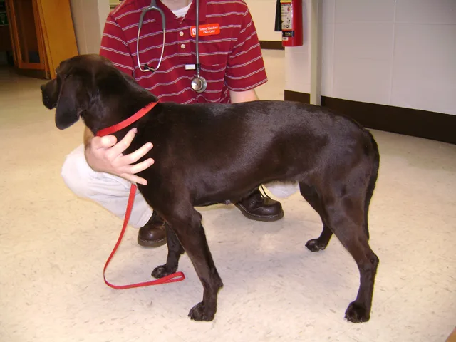
For waist palpation, both thumbs should be placed near the backbone with the fingers right behind the last rib (Figure 7). The width of the abdomen directly behind the last rib should be smaller than the width of the ribs and should slightly increase near the pelvic region (ie, hour-glass appearance).

FIGURE 7
Tail-Base Area Palpation
For tail-base area palpation, the thumb should be placed on one hip bone and all other fingers except the index finger on the opposite hip bone (Figure 8). The index finger should be used to palpate the dorsal spinous processes of the vertebrae in the region. Dogs with an ideal BCS of 3/5 or 4-5/9 should have a smooth contour in the palpated area, but bones can be felt under a thin layer of fat. The hip bones and dorsal spinous processes become more prominent and may protrude in underweight dogs and not be palpable as a dog becomes obese. Obese dogs can also develop rolls of fat in the tail-base region that give the appearance of a dimple.

FIGURE 8
Overweight vs Obese
In the United States an estimated 24% to 44% of dogs are overweight or obese, and ≈50% of dogs between 5 and 10 years of age are obese. Dogs 10% to 20% above ideal body weight are considered overweight. Obesity is the excessive accumulation of adipose tissue in the body and occurs when dogs are >20% above ideal body weight. Obesity is the most common nutritional disorder detected in dogs.
Muscle Condition Score
Muscle condition score (MCS) is commonly used to evaluate body muscle mass. MCS is assessed through visualization and palpation of the spine (ie, epaxial), scapulae, skull, and ilial wings. Epaxial muscle loss is typically the first indication of a decrease in muscle mass; muscle loss at other anatomic sites may be more variable. MCS is graded as normal, mild, moderate, or severe loss. Importantly, BCS and MCS are not directly related, as patients can have significant muscle loss despite an overweight body condition. Conversely, patients can have a low BCS despite a normal MCS. Palpation of muscle loss is particularly important in patients that are overweight or experiencing mild muscle wasting. Muscle loss associated with pathology is referred to as cachexia, and muscle loss associated with aging in the absence of disease is referred to as sarcopenia; both may occur concurrently.4
AAHA and WSAVA recommend MCS be performed as part of a nutritional assessment on all dogs and cats at every clinic visit.5,6