Arterial and venous blood gas samples can be used to assess acid-base status, electrolyte levels, and respiratory function. Arterial samples are the standard for measuring oxygenation (partial pressure of oxygen of arterial blood gas [PaO2]) and ventilation (partial pressure of carbon dioxide of arterial blood gas [PaCO2]).1
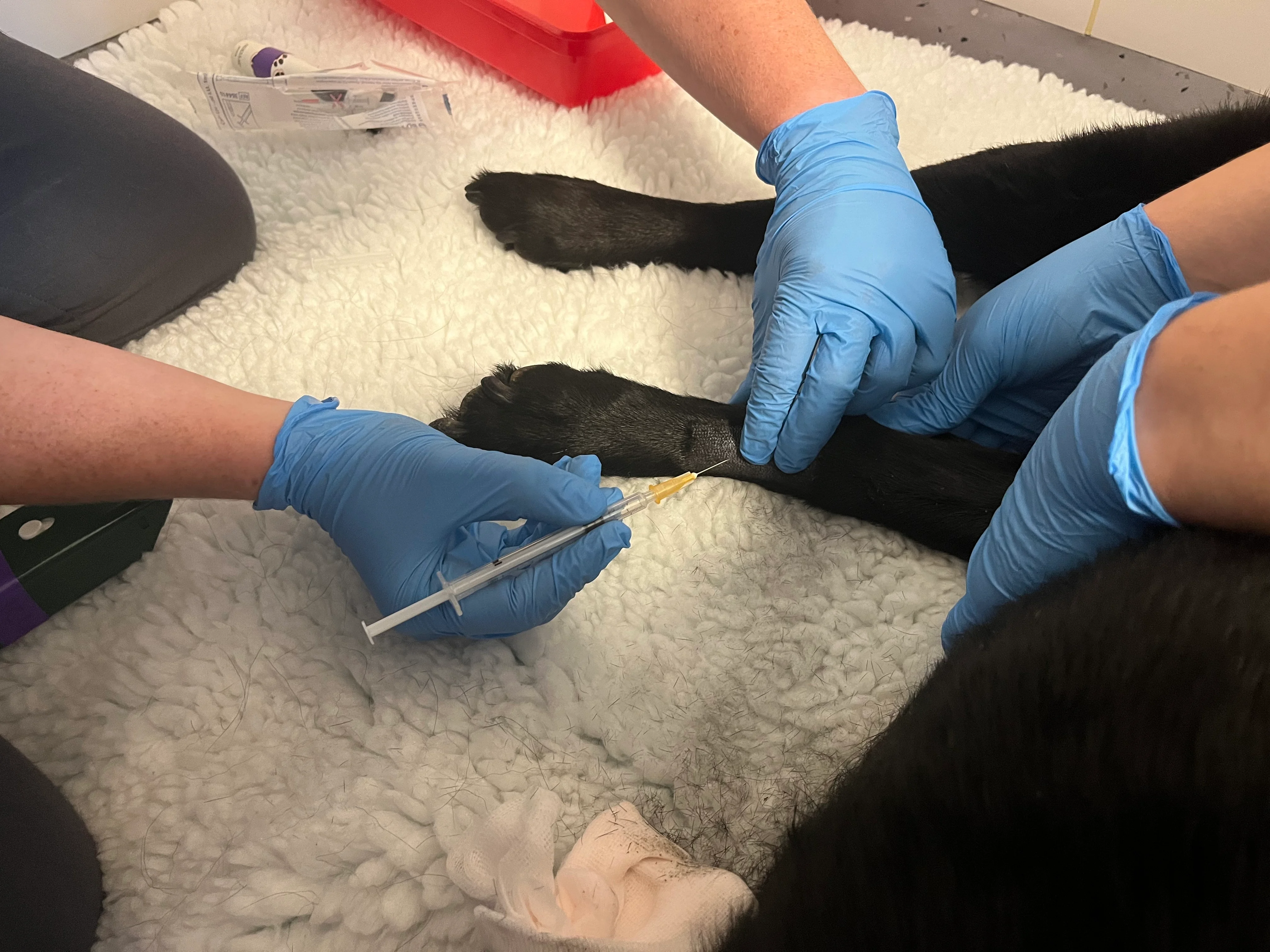
Samples for arterial blood gases can be obtained from several sites, including the dorsal pedal, femoral, lingual, and coccygeal arteries. The dorsal pedal artery is the preferred sampling site because it is easy to access and a pressure bandage to minimize bleeding can be applied following sample collection.2
Arterial blood samples should be collected in a heparinized syringe to prevent the sample from clotting. Purpose-made heparinized blood gas syringes that allow for automatic syringe filling with the arterial pulse are available (Figure 1). Standard syringes can be used and filled with heparin; however, heparin should be maintained at <4% of the total blood volume to avoid excessive sample dilution.3

FIGURE 1 A purpose-madeheparinized blood gas syringe
Following collection with a narrow-bore needle, air should be expelled from the syringe, and the sample should be capped immediately to avoid exposure to atmospheric air and subsequent sample contamination (Figure 2). Air bubbles should also be removed from the sample to reduce analytical error.

FIGURE 2 A sample capped to avoid air exposure and contamination
Complications of arterial sampling are rare but include pain, hemorrhage, hematoma formation, thrombosis, and infection at the sampling site.1 Arterial sampling is contraindicated in patients with thrombocytopenia or coagulopathy.
Step-by-Step: Blood Sampling of the Dorsal Pedal Artery4
What You Will Need
Examination gloves
Hair clippers
Diluted chlorhexidine solution (2% or 5%)
Small-bore needle (25 or 27 gauge)
Heparinized blood gas syringe
Pressure bandage materials

Step 1: Restrain the Patient
While wearing examination gloves, restrain the patient in lateral recumbency with the pelvic limb used for sampling placed on the dependent side.
Author Insight
Sedation is typically not necessary; however, butorphanol can be considered if deemed safe in a patient that is potentially hypoxemic.
A team member should hold the distal thoracic limbs with one hand and place their forearm over the patient’s neck to control the head, ensuring normal breathing is not restricted. The other hand should grasp the patient’s Achilles tendon above the hock to restrain the leg that will be used for sampling.

Step 2: Palpate the Pulse
Palpate the pulse on the dorsal medial aspect of the dependent limb.
Step 3: Prepare the Site
Clip the hair, and use a diluted chlorhexidine solution to aseptically prepare the arterial sampling site.

Step 4: Insert the Needle
Palpate and localize the arterial pulse using a finger on your nondominant hand. Hold a heparinized blood gas syringe between the thumb and first fingers of your dominant hand (similar to holding a pen), and advance the needle into the vessel at a 45-degree angle directly below the palpated arterial pulse.

Author Insight
The force of the arterial pulse should fill the syringe passively.

Step 5: Remove the Needle
After an adequate sample is obtained, remove the needle, and apply direct pressure with a pressure bandage over the arterial puncture site for 5 minutes. Closely monitor the site for bleeding.

Step 6: Secure the Sample
Ensure all air is expressed from the syringe immediately after sample collection by holding the syringe vertically with the needle pointing up and tapping gently with a finger. Quickly cap the sample to avoid exposure to atmospheric air.

Author Insight
Samples should be analyzed immediately after collection. If immediate analysis is not possible, samples should be capped and stored on ice.5
Sampling Errors
Exposure to room air: Samples that are exposed to room air or contain air bubbles quickly equilibrate with the environmental partial pressures of oxygen and carbon dioxide, causing an artificial increase in PaO2 and a decrease in PaCO2 of the arterial sample.
Delay in sample analysis: Prolonged pre-analytical times may result in increased lactic acid concentration caused by anaerobic metabolism, which causes a decreased base excess and reduction in pH of the sample.
Interpretation of Arterial Blood Gas Results
Hypoxemia is defined as PaO2 <80 mm Hg or oxygen saturation (SpO2) <95%. Hypoventilation is defined as PaCO2 >45 mm Hg or partial pressure of carbon dioxide in venous blood (PvCO2) >55 mm Hg.6
The ratio of PaO2 to fraction of inspired oxygen (FiO2; P:F ratio) and the alveolar-arterial (A-a) gradient can assess oxygenation. Only arterial samples can be used to assess oxygenation. Conversely, both arterial and venous samples can be used to assess ventilation. PaCO2 from arterial samples is ≈3 to 6 mm Hg lower than in venous samples (PvCO2).7
P:F ratio
The P:F ratio can help determine the adequacy of oxygenation (which is particularly useful for patients receiving oxygen supplementation) and oxygen supplementation requirements.6
In general, PaO2 should be ≈5 times FiO2. Room air is 21% oxygen (0.21). A patient without lung disease breathing room air would therefore have a PaO2 of ≈100 mm Hg.
A normal P:F ratio is ≈500. For example, in a healthy patient breathing room air, the P:F ratio is 476: 100/0.21 = 476 mm Hg.
P:F ratio ≤200 mm Hg suggests significant pulmonary disease, defined as Veterinary Acute Respiratory Distress Syndrome (ie, VetARDS); P:F ratio ≤300 mm Hg is defined as Veterinary Acute Lung Injury (VetALI). For example, in a patient with pulmonary disease and PaO2 of 78 mm Hg breathing 30% oxygen via nasal cannula, the P:F ratio is 260: 78/0.3 = 260 mm Hg, which is consistent with VetALI.
The P:F ratio is most useful for evaluation of oxygenation in patients receiving supplemental oxygen. The A-a gradient or 120 rule (see The 120 Rule) should be used to evaluate oxygenation in patients breathing room air.
Alveolar-Arterial Gradient
A-a gradient is a measure of the difference between the concentration of oxygen in alveoli (PAO2) and the concentration of oxygen in arterial blood (PaO2).6 This gradient uses the following formula to help establish whether hypoxemia is due to ventilation failure (ie, hypoventilation) or pulmonary disease8:
PAO2 − PaO2
To calculate the A-a gradient, PAO2 must first be calculated using the alveolar gas equation:
PAO2 = [FiO2 × (Pb-PH20)] – PaCO2/RQ
FiO2 is expressed as a decimal; Pb (atmospheric pressure at sea level) is 760 mm Hg; PH20 (partial pressure of water) is 47 mm Hg; and RQ (respiratory quotient) is generally 0.8 but can vary based on diet and metabolism. For patients living at sea level and breathing room air, the formula can be simplified to:
PAO2 = 150 – (PaCO2/0.8)
For patients breathing room air, a normal A-a gradient should be <10 mm Hg; values >20 mm Hg indicate decreased oxygenation efficiency.
The 120 Rule
PaCO2 + PaO2 (ie, the 120 rule) can be used to assess lung function in patients breathing room air and assesses oxygenation alongside ventilation, as a change in one value can affect the other.6
In healthy patients, PaCO2 added to PaO2 should equal at least 120. For example, a patient with a normal PaCO2 of 40 mm Hg and a minimum normal PaO2 of 80 mm Hg will have a PaCO2 + PaO2 value of120.
The 120 rule can also help determine whether hypoxemia is due to hypoventilation alone or both hypoventilation and lung dysfunction. For example, if a patient has an increased PaCO2 of 65 mm Hg and a PaO2 of 55 mm Hg, then PaCO2 + PaO2 = 120, which indicates hypoxemia is due to hypoventilation alone. If the patient instead has a PaCO2 of 65 mm Hg and a PaO2 of 35 mm Hg, then PaCO2 + PaO2 = 100, which indicates hypoxemia is due to lung dysfunction in addition to hypoventilation.
Example of an Arterial Blood Gas
The patient in Table 1 is breathing room air; FiO2 is 21%.
The simplified formula for PAO2 is 150 − (21.6/0.8), which equals PAO2 of 123. The A-a gradient is thus 123 − 48.9 = 74.1. This result indicates severe venous admixture or markedly decreased oxygen deficiency. Using the 120 rule, the PaCO2 + PaO2 value is 70.5, which is markedly below 120 and indicates severe lung dysfunction.