Common Tibial Plateau-Leveling Osteotomy Complications
Dominique Hemmings, DVM, Tuskegee University
Selena Tinga, DVM, PhD, DACVS-SA, The Ohio State University
The cranial cruciate ligament (CrCL) resists cranial tibial translation, internal tibial rotation, and stifle hyperextension.1 Rupture of the CrCL (CrCLR) is the most common cause of hindlimb lameness in dogs,1 often resulting in instability, meniscal tearing, and osteoarthritis. CrCL degeneration is caused by a combination of factors, including age, obesity, trauma, genetics, and abnormal bony morphology.1
Complete CrCLR can typically be diagnosed via palpation (positive cranial drawer or tibial thrust), although early or partial tears are more challenging to diagnose.1 Radiography should be performed to identify findings supportive of CrCLR (eg, stifle joint effusion, osteoarthritis, cranial tibial subluxation), to rule out other causes of pain or instability (eg, fractures, neoplasia), and for surgical planning.1
Treatment options for CrCLR include osteotomy-based procedures, extracapsular suture procedures, and nonsurgical management. A survey suggested that surgeons prefer tibial plateau-leveling osteotomy (TPLO) for treatment of most cases of CrCLR in dogs weighing >33 lb (15 kg).2-7 Several studies report an equal to superior outcome for TPLO as compared with extracapsular suture procedures and other osteotomy procedures.3-7 For example, some studies have shown TPLO to have a quicker return to normal weight bearing, higher pet owner satisfaction, and slower progression of osteoarthritis.3-7 However, 10% to 34% of dogs treated with TPLO develop a complication, with up to 4% requiring revision surgery.7 Complications may arise from inappropriate candidate selection, imperfect surgical technique, or poor owner compliance, but complications are also an inherent risk of surgery in any patient.
Common TPLO Complications
Surgical Site Infection
The incidence of surgical site infection (SSI) after TPLO ranges from 1.3% to 25.6%, with a wide reported range secondary to variable definitions of SSI risk factors and methodology.8-12 SSI can occur in superficial and/or deep tissues and includes the subcategories of soft tissue infection, implant-associated infection, osteomyelitis, or septic arthritis. A large review reported the incidence of SSI subcategories occurring after TPLO by taking case numbers from numerous primary studies; wound complications occurred in 7.8% of TPLO cases, implant-associated infection occurred in 3.4%, osteomyelitis occurred in 0.6%, and septic arthritis occurred in 0.8%.6
Reported risk factors for development of post-TPLO SSI include German shepherd breed, heavier body weight, undergoing a meniscectomy, inexperienced surgeon, and prolonged duration of surgery and anesthesia.8-10 One study reported a nonsignificant trend toward increased SSI rate after TPLO in dogs with dermatitis8; thus, the authors recommend giving consideration to surgical delay and controlling dermatitis prior to elective orthopedic surgery, particularly when dermatitis is severe or within the surgical site. In addition to sterile technique, the risk for SSI is likely mitigated by meticulous tissue handling, accurate wound closure, minimizing the duration of surgery and anesthesia, copious lavage, and perioperative administration of a first-generation cephalosporin antibiotic (eg, cefazolin).8,9 One study documented a significant reduction in SSI rate from 8.5% to 1.3% after implementation of a strict infection control protocol that included use of an adhesive iodine-impregnated drape during surgery, single use gloves at all times when handling dogs, and an Elizabethan collar, in addition to multiple additional efforts.11
The use of perioperative antibiotics is supported, but there is conflicting evidence regarding the use of postoperative antibiotics, and the potential protective effects must be weighed against the risk for developing bacterial drug resistance.8-10 If an SSI occurs, immediate and aggressive wound management, bacterial culture, and antibiotic therapy are all recommended to control the infection while the bone heals. Even superficial soft tissue infections can progress to implant-associated infections and osteomyelitis, especially if not treated appropriately, and can result in delayed union, nonunion, or persistent infection (Figures 1 and 2).7 Treatment for an implant-associated infection, osteomyelitis, or septic arthritis requires long-term antibiotic therapy, possible surgical flush, debridement, and/or local antimicrobial therapies, as well as implant removal in many cases, once bone healing is confirmed.13
In rare cases, infection cannot be controlled, bone healing cannot be achieved, and amputation or euthanasia is required.


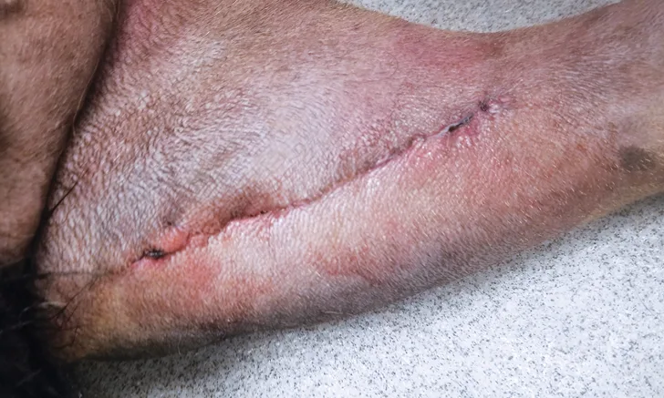
A 5-year-old neutered male Rottweiler that developed a deep surgical site infection 5 days after TPLO. Prior to removal of intradermal sutures (A), thick serosanguinous discharge was easily expelled from the incision in multiple locations (inset). After removal of intradermal sutures (B), the TPLO plate was immediately visible, indicating dehiscence of the fascial closure, and the tissues appeared inflamed and were coated with a thick mucoid film. The wound was managed as an open wound for 4 days then closed once tissues appeared healthy, and the implant was not removed; the patient remained on oral culture-based antibiotics until healing of the osteotomy (delayed union). This dog did not develop osteomyelitis and had no lameness at the last follow-up.

Radiographs from an 8-year-old spayed Rottweiler that underwent TPLO and was diagnosed with a superficial SSI 2 weeks postoperatively at another hospital. The SSI was treated with a 10-day course of antibiotics. The dog was presented to The Ohio State University Veterinary Hospital 6 weeks after surgery for recurrent lameness; the incision was healed, but osteomyelitis was confirmed on radiographs and fine-needle aspirate and cytology. Culture-based antibiotics were prescribed, but the infection did not resolve, the lameness was persistent, and the osteotomy became a nonunion. The patient was euthanized after developing a T3-L3 myelopathy suspected to be related to systemic infection.
Residual Instability
Cranial-caudal stifle instability is present postoperatively in one-third of TPLO-treated patients.14 Though the majority of dogs with postoperative instability are nonclinical, even nonclinical residual instability may result in a reduced long-term outcome. A more severe instability known as pivot shift, which involves cranial tibial subluxation coupled with a sudden lateral motion of the stifle during weight bearing, occurs in up to 3% of cases.7,12 The cause of residual instability has been hypothesized to be related to meniscectomy (or meniscal release) or incomplete plateau leveling.12,14 Incorrect osteotomy position (eg, osteotomy is positioned distally; Figure 3) or plateau rock-back can affect success in achieving or maintaining plateau leveling and therefore may affect stifle stability.1 In some cases, stifle instability after TPLO (including pivot shift) may resolve with time,8 likely due to improved muscular strength, which may support the hypothesis that some degree of instability can occur due to muscle weakness and further support the recommendation for postoperative physical therapy.

Immediate postoperative radiographs from a 2-year-old spayed medium-size crossbreed dog showing an inappropriately distally positioned TPLO. Distalizing the TPLO reduces the leveling achieved with planned rotation, leaves a narrow tibial crest (arrow), and positions the osteotomy in diaphyseal bone (slower to heal than metaphyseal bone). Also notable is the cranial position of the distal jig pin hole, which may predispose the patient to tibial diaphyseal fracture. This osteotomy position can be compared with that shown in Figure 4, in which the osteotomy position and resultant crest shape are appropriate.
Medial Meniscal Tears
Meniscal pathology causes lameness and progression of osteoarthritis; therefore, intra-articular examination at the time of TPLO is necessary for the diagnosis and treatment of concurrent meniscal pathology. In the months following TPLO, postoperative meniscal tears are diagnosed in 1.8% to 10.5% of cases in which the meniscus was classified as normal and left untreated at the time of TPLO.6,15,16 Some of these cases likely represent meniscal tears that were present but not identified at the time of original surgery. The sensitivity of detecting meniscal tears can be increased by using arthroscopy (vs arthrotomy) and by using a stifle distractor and meniscal probe during joint examination.1,16,17 Development of a postoperative meniscal tear is likely related to the presence of residual stifle joint instability and often results in persistent lameness, requiring an additional procedure for meniscal debridement.
Patellar Tendinosis
Patellar tendon thickening (Figure 4) is a benign process that occurs in 80% to 100% of dogs after TPLO.18 In up to 7% of cases, this thickening is associated with pain and lameness (patellar tendinosis).18 Patellar tendinosis usually responds to NSAIDs and rest, followed by gradual return to activity,18 with anecdotal evidence also supporting the use of shockwave therapy and physical rehabilitation therapy.

Radiographs from a 7-year-old spayed golden retriever presented with recurrent lameness 3 months after TPLO. A moderate weight-bearing lameness and pain on palpation of the cranial stifle/patellar tendon was identified on examination of the operated limb. Radiographs revealed thickening of the patellar tendon (solid arrows) and an apical patellar fracture (dashed arrow). Lameness resolved with rest, NSAID therapy, and shockwave therapy.
Intra-Articular Screw Placement
Intra-articular screw placement likely leads to persistent pain and hastened osteoarthritis development if not addressed immediately. The incidence of intra-articular screw placement during TPLO ranges from <0.1% to 7.1%.6,12,19,20 In one study comparing the use of locking and nonlocking TPLO plates, nonlocking TPLO plates were associated with an increased risk for intra-articular screw placement; this is likely because locking TPLO plates are typically precontoured with a screw trajectory designed to minimize the risk of intra-articular or intra-osteotomy screw placement.20 However, poor plate positioning, intraoperative plate contouring, or cross-threading can affect screw trajectory and result in intra-articular screw placement when using locking plates (Figure 5).20 Postoperative radiographs must be scrutinized for intra-articular screws, and offending screws must be immediately redirected or shortened to prevent the long-term effects of this complication.

Radiographs from a 5-year-old neutered male Bernese mountain dog with persistent lameness 2 months following TPLO. Radiographs revealed the proximal-most screw violating the joint space (arrow). This is best visualized on the third (oblique) view and was not identified on immediate postoperative radiographs. The locking plate is designed to reduce the risk of intra-articular screw placement, but this plate was contoured intraoperatively to accommodate for excessive medial buttress, which resulted in a screw trajectory directed toward the joint space.
Uncommon Complications
The following complications are uncommon but can be catastrophic and therefore warrant individual discussion. Poor surgical technique will increase the incidence of these complications.
Plateau Rock-Back
Rock-back (ie, loss of plateau leveling) results from failure—sometimes catastrophic—of the plate, screws, and/or plateau segment. Some studies report more loss of osteotomy reduction in the postoperative period when nonlocking constructs are used as compared with locking constructs.21 Rock-back can affect long-term outcome if it results in recurrent stifle instability. Implant or bone failure can occur with use of either nonlocking or locking constructs in cases of poor surgical technique (eg, poor osteotomy position, incomplete osteotomy compression, improper plate/screw position or application) or incomplete postoperative activity restriction and can havecatastrophic consequences.
Tibial Tuberosity Fracture
The risk for tibial tuberosity fracture may be increased by an osteotomy position that results in a narrow crest (Figure 3), by bilateral simultaneous TPLO procedures, or by other factors that either decrease the strength of the patellar tendon’s anchor point or increase the pull of the patellar tendon.7,22 Many cases do not require intervention, although surgical stabilization may be required if the fragment is unstable.
Fibular Fracture
Inadvertent drilling of the fibula during TPLO increases the risk for fibular fracture 10-fold. Although it is suspected that the risk for fracture is higher when the fibular drill hole is left unfilled—and therefore it is recommended to fill the hole—this was not proven statistically (likely a type II statistical error).23 Increased body weight is also a risk factor for fibular fracture after TPLO.23 Fibular fracture eliminates the fibula’s splinting function, which likely aids in stabilizing the osteotomy and, therefore, fibular fractures may increase the incidence and degree of rock-back.23
Patellar Luxation
Patellar luxation occurs in <1% of cases following TPLO, although in one study, the majority of postoperative patellar luxations required revision surgery.7,15 The theorized causes of patellar luxation as a complication of TPLO include muscle atrophy, closure of the medial retinaculum under too much or too little tension, severe joint effusion after surgery, and creation of tibial malalignment.15
Other Complications
Additional complications to consider include anesthetic complications, minor incisional complications (eg, minor dehiscence, seroma, suture reaction), intraoperative hemorrhage (arterial), delayed/nonunion, implant failure, tibial diaphyseal fracture, creation of angular deformity, collateral ligament or patellar tendon trauma, and implant-associated sarcomas.7,15,24
Conclusion
Preventing complications during recovery depends on both preoperative and intraoperative decision making, along with owner education and compliance. Owners must be instructed to keep Elizabethan collars on their pet until the incision is healed. For ≈8 weeks following surgery, or until radiographic healing is demonstrated, patient activity should be strictly controlled; no concussive activity or free roaming should be allowed, but gradually increasing duration of leashed walking and other controlled strengthening exercises is important to promote muscular recovery and bone healing. Adhering to these strict guidelines should mitigate the risk for incisional complications, implant and bone failure, and delayed healing.
CrCL = cranial cruciate ligament, CrCLR = cranial cruciate ligament rupture, SSI = surgical site infection, TPLO = tibial plateau-leveling osteotomy