Skills for Technicians: Urine Sample Collection & Analysis
Kirk Ryan, DVM, DACVIM, Louisiana State University
Urinalysis is a common diagnostic test in veterinary medicine used to help identify a wide range of conditions such as kidney disease, urinary tract infection, and diabetes.
Home Urine Collection
Voided urine samples, which many clients can obtain at home, are generally not intended for culture because of the potential for contamination, but they are adequate for simple urinalysis. Small amounts of urine can be used for urinalysis, but a minimum 8 mL sample is preferred to maximize urine sediment examination.1
For cats, place nonabsorbent plastic beads or polished gravel in the litter box. After the cat urinates, the urine can be aspirated into a syringe or decanted into a container and taken to the practice. For dogs, a midstream urine sample can be collected in a clean or sterile container. Suitable sample collection containers provided by the practice are preferred over improvised home solutions (eg, disposable, easily sealed containers such as those that hold margarine or yogurt).
Clinical Urine Collection
Cystocentesis is the preferred route of sample collection in most cases. Most cats and small dogs are restrained in lateral recumbency to permit bladder palpation. Retract the hind limbs for greatest access to the caudal abdomen for palpation. Then, stabilize the bladder against the body wall, puncture it percutaneously, and withdraw urine with a needle and syringe.
Larger dogs are placed in dorsal recumbency and may require 2 team members for safe restraint. Estimate the bladder’s approximate location by drawing an imaginary X between the caudal 4 mammae. For males, retract the penis and prepuce laterally. Advance the needle percutaneously into the bladder on midline to avoid pain and iatrogenic hemorrhage (eg, when the needle is advanced through muscle rather than the avascular and firm connective tissue of the linea alba). Use ultrasound to locate the bladder and assess urine volume prior to sampling. With practice, cystocentesis may be performed under ultrasound guidance to visualize the needle’s advancement into the bladder. Cystocentesis may also be performed in some large-breed dogs with the patient restrained in a comfortable standing position.
Collecting urine via a urinary catheter is sometimes desirable. Catheters placed using sterile techniques permit collection of a sample suitable for culture.
Generally, place sterile urine samples in labeled, plain glass, red-top tubes for transport to the laboratory. A good practice following collection of all sterile urine samples is replacing the sampling needle with another sterile needle before transferring a few urine drops into a separate sterile tube for culture. Perform a urinalysis within 2 hours of urine collection; if this is not possible, store the sample in a refrigerator for later analysis at room temperature.
Urine Sample Collection & Analysis

Figure 1
Cystocentesis is performed on a male dog restrained in dorsal recumbency. Note the retraction of the prepuce to permit comfortable introduction of the sampling needle. Photo courtesy of Kirk Ryan, DVM
Expert Tip
If urine is not obtained on the first attempt, avoid redirecting the needle, which may inadvertently lacerate abdominal organs. Similarly, if patient restraint is inadequate and the animal moves suddenly, the needle should be immediately withdrawn. Cystocentesis complications have rarely been reported but can include puncture or laceration of the aorta or bladder and may result in severe hemorrhage or urine leakage into the abdomen. Vasovagal syncope (ie, fainting or collapsing) may occur from autonomic stimulation of the bladder but is very rare.2
Laboratory Analysis
A complete urinalysis is comprised of several components.
Record urine color and subjectively assess turbidity as clear, hazy, or cloudy.
Measure urine specific gravity (ie, urine concentrating ability) using a handheld refractometer, which relates the refractive index of liquids to their concentration of dissolved solids. Place a drop of urine on the refractometer lens, holding it toward light to permit reading of the refractive index on a standardized scale where distilled water has a specific gravity of 1.000.Dilute urine has a urine specific gravity of ≤ 1.007. Concentrated urine has a specific gravity of ≥ 1.013. (Very concentrated urine has a specific gravity of ≥ 1.030 in the dog and ≥ 1.035 in the cat). Urine with a specific gravity of 1.008 to 1.012 is considered isosthenuric (ie, having the same concentration as normal plasma).3
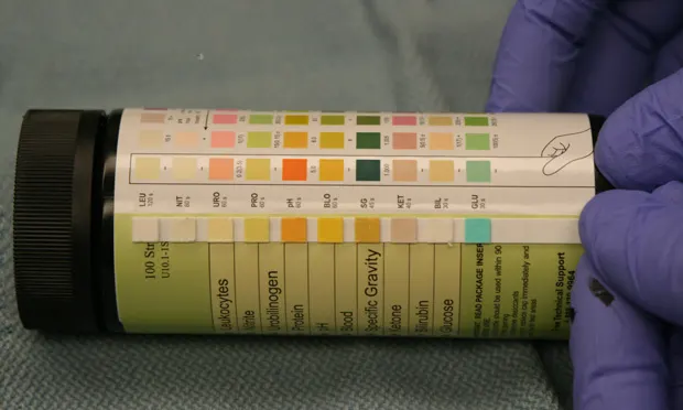
Measure urine pH and relative amounts of urine protein, glucose, ketones, bilirubin, urobilinogen, blood, leukocyte, and nitrite concentrations using laboratory reagent strips that react biochemically with urine to produce a color change. Expose the test strips to urine following manufacturer guidelines and use a color-coded legend to interpret results.
Expert Tip
Because urine dipstick values depend on detection of reagent strip color change, substances that alter urine color (eg, bilirubin, hemoglobin, myoglobin) may interfere with interpretation (ie, colorimetric interference).
Examine the urine sediment microscopically in conjunction with urine dipstick biochemical assays. Any urinalysis performed without such examination is considered incomplete. Examine urine sediment for the presence of red and white blood cells, renal tubular casts, bacteria, and crystals. These urine sediment components are quantified by a specific number per high power field (eg, 10-20/hpf), or more qualitatively as rare, occasional, many, or too numerous to count (TNTC).
To obtain urine sediment for examination, place a sample tube containing urine (ideally, 5 mL) in a balanced centrifuge and spin at 400 G for 5 minutes. Many veterinary practice centrifuges have a preset speed- and time-control setting for urinalysis. During centrifugation, solid materials suspended in the urine will gravitate into a pellet at the bottom of the tube. Allow the centrifuge to stop spinning naturally; engaging the centrifuge brake may result in the pellet’s resuspension. The pellet contains the urine sediment, which is collected via pipette or by decanting the supernatant fluid.
Resuspend the pellet in the small amount of fluid remaining in the tube. Place a drop of sediment on a clean glass microscope slide and cover with a coverslip. After scanning the slide at low magnification (10x), examine at least 10 fields of view at 40x magnification to assure representative reporting. Lower the condenser on the microscope to increase the sediment contrast. Commercial stains (ie, Sedi-Stain) can aid visualization, but unstained evaluation in proper lighting is usually adequate.
Conclusion
Collecting urine and performing a urinalysis is a fundamental skill for veterinary technicians. This crucial diagnostic test is often overlooked because of the perceived inconvenience of sampling, but urinalysis is a key health and hydration indicator that can provide important clues to many disease diagnoses.