A Feline Challenge
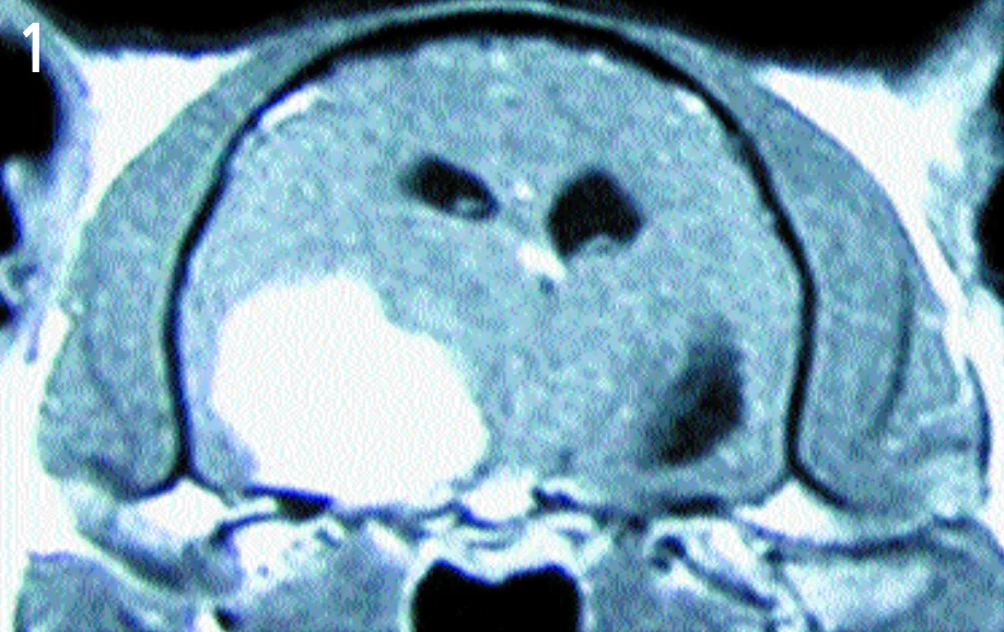
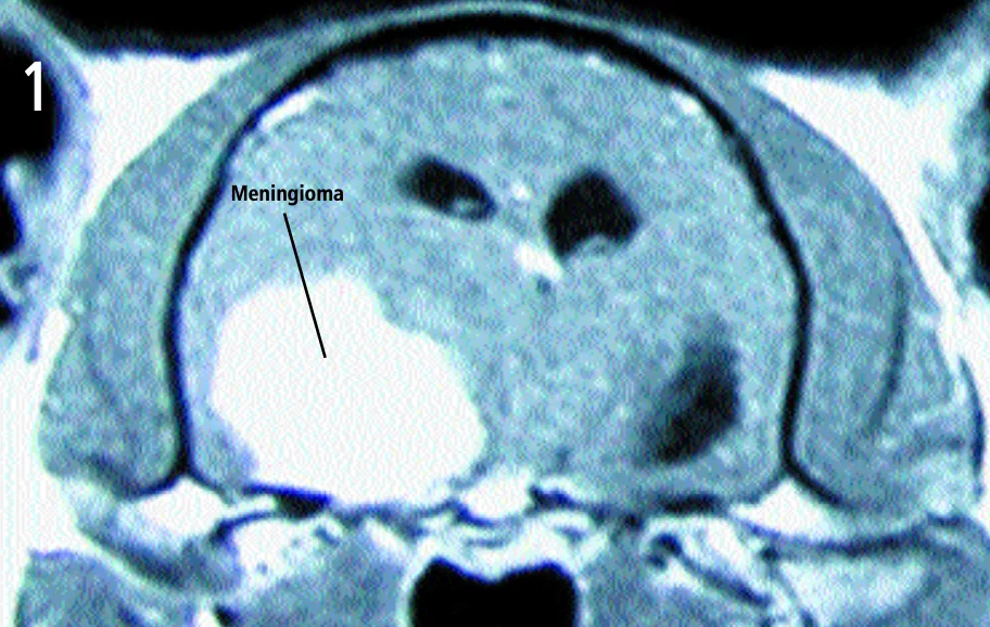
Transaxial view—T1-weighted, contrast-enhanced magnetic resonance image
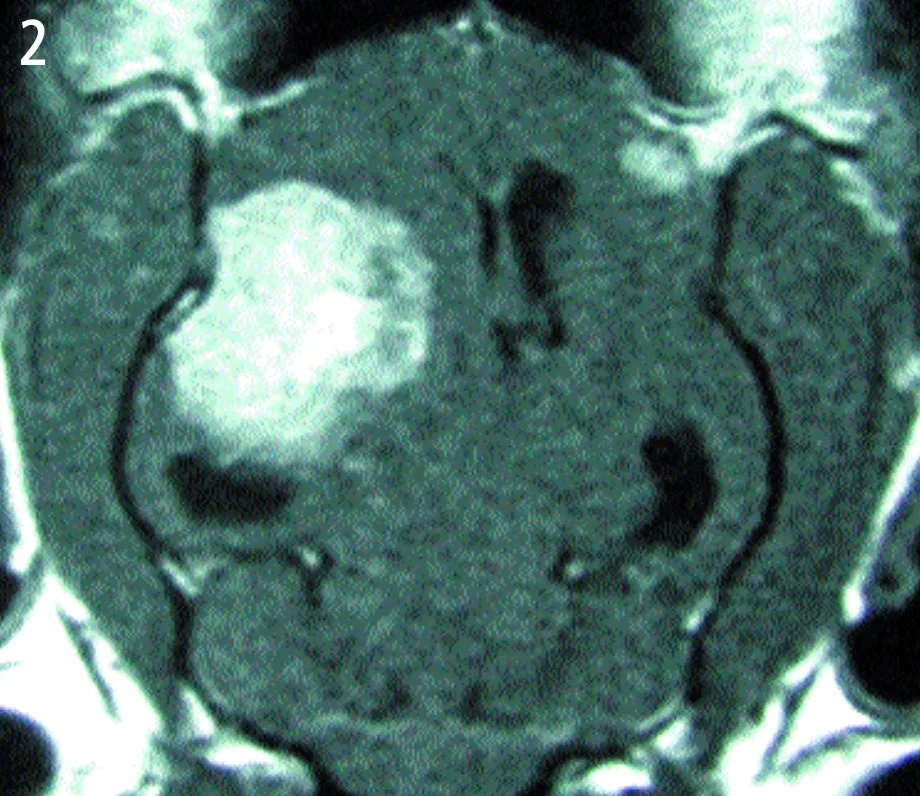
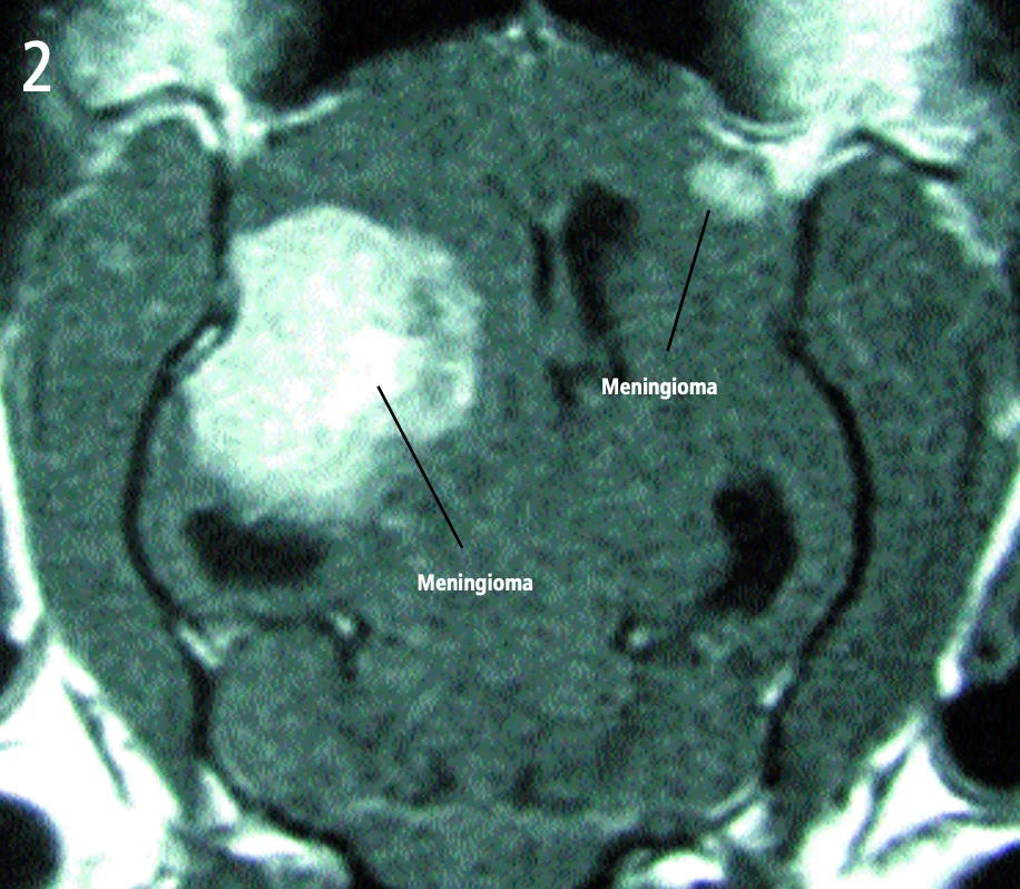
Dorsal view—T1-weighted, contrast-enhanced magnetic resonance image
History. A 10-year-old, female, spayed domestic shorthair cat is presented to the hospital. The cat has a 3-week history of circling to the left and occasional aggression toward the owner.
Examination. Other than being overweight, no abnormalities are found on physical examination. Abnormalities identified on neurologic examination include decreased menace reaction on the right side, head turn to the left, tendency to circle to the left, and decreased facial sensation on the right side. The patient also tends to ignore environmental cues such as sounds, food, and skin pinch, when applied to the right side. Although the cat walks in a circle, gait is normal.
Table: Laboratory Work
ALT = alanine aminotransferase; BUN = blood urea nitrogen; HCT = hematocrit; WBC = white blood cell
Ask Yourself ...
What is the principal abnormality apparent on the images?
Are there any other obvious lesions?
What is the most logical course of therapy for this problem?
What is the prognosis?
A large, left-sided cerebral mass, evident on both images.
The mass is uniformly contrast-enhancing on the MRI, appears to have a broad-based attachment to the skull, and has distinct margins-all of which are characteristic of intracranial meningiomas. The most likely diagnosis is meningioma. A smaller, right-sided mass can be seen on the dorsal image. This mass also has characteristic features of meningioma. Both masses were removed and confirmed histologically as meningiomas. The cat made a full recovery.
The neurologic deficits in this cat point to a lesion in the left forebrai (cerebrum/diencephalon). Patients with unilateral forebrain lesions tend to circle toward the side of the lesion and demonstrate sensory deficits (e.g. conscious proprioception, vision, facial sensation) opposite the lesion side. The phenomenon of ignoring environmental cues on the side opposite a focal forebrain lesion is termed hemineglect (or hemi-inattention) syndrome. Since most sensory input is normally interpreted on the cerebral hemisphere on the opposite side from which the stimulus is applied, a left cerebral mass results in the patient ignoring the right side of his/her environment.1
Transaxial view—T1-weighted, contrast-enhanced magnetic resonance image
Dorsal view—T1-weighted, contrast-enhanced magnetic resonance image
There are many possible causes for a focal forebrain lesion in an older cat, but a brain tumor is by far the most likely, and meningioma is by far the most common feline brain tumor.2 Multiple intracranial meningiomas occur in cats relatively infrequently. Most cats with intracranial meningiomas recover quickly after surgical removal. Median survival is about 2 years with surgery alone.<sup1-3 sup>
DID YOU ANSWER ...
A large, left-sided cerebral mass
A smaller, right-sided cerebral mass
Surgical removal
Good prognosis