Weakness & Collapse in a Labrador Retriever
History. The dog was current on vaccines and receiving monthly heartworm prophylaxis. Approximately 3 weeks before development of the current signs, the dog had a bout of gastroenteritis associated with lethargy. She had recovered uneventfully after receiving intravenous fluids and a bland diet. Forty-eight hours before presentation, the dog again developed lethargy and anorexia. Vomiting and weakness also occurred, and the dog was presented for evaluation at the family veterinarian and was referred to Tufts for ongoing evaluation and treatment.
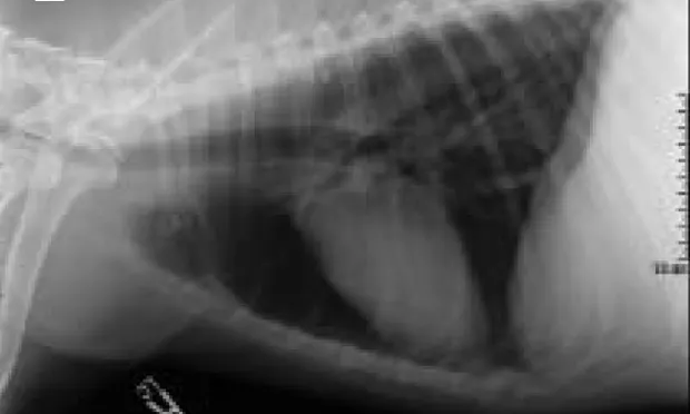
Physical Examination. The dog was weak and needed to be gurneyed to the treatment room. Heart rate was 75 bpm, respiratory rate was 24 breaths/min, rectal temperature was 100.7 °F, and pulse quality was weak. Body weight was 35 kg (77 pounds). Mucous membranes were pale, and capillary refill time was prolonged at 3 seconds. An ECG was obtained (Figure 1). Pertinent point-of-care blood analysis findings are listed in the Table. Urine was collected via cystocentesis; urine specific gravity was 1.018, dipstick findings were unremarkable, and sediment was inactive. A lateral chest radiograph was taken (Figure 2).
ASK YOURSELF ...• What is the ECG abnormality?• What abnormality is present on the chest radiograph?• What are the most appropriate initial diagnostic and treatment plans?• What is the most likely diagnosis?• What is the long-term prognosis?
Diagnosis: Addison's disease
Laboratory findings that are consistent with Addison's disease include lymphocytosis and eosinophilia (lack of a stress leukogram), hyponatremia, hyperkalemia, hypercalcemia, and hypocholesterolemia. Anemia or hemoconcentration, melena, azotemia, and hypoalbuminemia may also be present. Thoracic radiographs often document hypovolemia and may show a gas-filled esophagus consistent with a megaesophagus.
Addison's disease is very rare in cats and is definitively diagnosed by an ACTH-stimulation test documenting an inadequate (usually undetectable-e.g., < 0.2 µg/dl (pre) and < 0.2 µg/dl (post) response to ACTH. Other diseases that may mimic clinical or laboratory findings of Addison's disease include whipworm infection or cavitary effusions, specifically pericardial effusion resulting in development of large volumes of ascites and decreased circulating volumes.
TreatmentSunny was treated with rapid infusion of 2.5 liters of saline over 30 minutes. Sinus rhythm was present on the ECG following the first liter. Based on the electrolyte abnormalities, an ACTH-stimulation test was performed, and the dog was treated with 24 mg IV dexamethasone. A high dose of glucocorticoids was chosen to address the patient's Addisonian crisis. The supplemental glucocorticoids were given before the results of the ACTH-stimulation test were obtained, as the index for suspicion of Addison's disease was high. Much lower doses of glucocorticoids are required for day-to-day stresses. Presample cortisol levels were less than 0.2 µg/dl, and postsample cortisol levels were less than 0.2 µg/dl, confirming the diagnosis.
The dog was admitted to intensive care and responded well to IV fluids. Azotemia resolved within 36 hours. She was given an injection of 77 mg DOCP and discharged with oral prednisone (2.5 mg per day) with additional prednisone to be given during times of crisis or stress. A reexamination with evaluation of electrolytes and BUN/creatinine was plannedfor 2 weeks later.
Did you answer ...
• The ECG (Figure 1; page 34) documented the absence of P waves, widened QRS complexes, and slight tenting of the T wave. These abnormalities are associated with hyperkalemia and reflect the effect of electrolyte abnormalities on the cardiac myocardium. It is important to note that the bradycardia in this dog is inconsistent with the other signs of cardiovascular collapse.• Interpretation of the lateral thoracic radiograph (Figure 2; page 34) reveals microcardia and a small caudal vena cava. The radiographic abnormalities are consistent with hypovolemia.• Diagnosis: Initial diagnostic testing should include a CBC, serum chemistry profile, urinalysis, and an ACTH-stimulation test.
Treatment: The most appropriate initial step is volume expansion with 0.9% saline. One or two 16- or 18-gauge catheters should be placed in a peripheral vessel and fluids infused until cardiovascular status improves and the ECG abnormalities are corrected. In general, this requires 60 to 100 ml/kg. In dogs with marked hyponatremia (e.g., < 125 mEq/dl), rapid correction of serum sodium may be associated with central pontine myelinolysis and subsequent neurologic decline. Too rapid correction of sodium deficits should be avoided. When Addison's disease is suspected, glucocorticoids should be administered at supraphysiologic doses (0.25 to 1.0 mg/kg dexamethasone). Dexamethasone is chosen as the replacement glucocorticoid instead of prednisone as dexamethasone will not interfere with the cortisol assay.
The clinical signs and laboratory findings are most consistent with Addison's disease (hypoadrenocorticism). This disease occurs most commonly in middle-aged, female dogs and is most often due to immune-mediated destruction of the adrenal gland, which results in deficiency of glucocorticoids/mineralocorticoids. Pituitary tumors, invasion or necrosis of the adrenal gland, or long-standing high-dose glucocorticoids may also affect adrenal function. Before the crisis, clinical signs may be vague or wax and wane.
The long-term prognosis for the dog is excellent. Dogs will require treatment with either daily (Florinef) or long-acting mineralocorticoids (DOCP); most dogs seem to do very well on long-acting therapy. Supplemental physiologic glucocorticoids (prednisone 0.1 to 0.2 mg/kg/day) should be available and the dose increased during times of stress.
WEAKNESS & COLLAPSE IN A LABRADOR RETRIEVER • Elizabeth Rozanski
Suggested Reading
Diagnosis and management of primary spontaneous hypoadrenocorticism (Addison's disease) in dogs. Kintzer P, Peterson M. Semin Vet Med Surg Small Anim Pract 9:148-152, 1994.Diagnosis and treatment of naturally occurring hypoadrenocorticism in 42 dogs. Melian C, Peterson M. J Small Anim Pract 37:268-275, 1996.Subcutaneous administration of desoxycorticosterone pivalate for the treatment of canine hypoadrenocorticism. McCabe MD, Feldman EC, Lynn RC, Kass PH. JAAHA 31:151-155, 1995.Treatment and long-term follow-up of 205 dogs with hypoadrenocorticism. Kintzer P, Peterson M. J Vet Intern Med 11:43-49, 1997.