Vulvar Discharge in the Bitch
Autumn P. Davidson, DVM, MS, DACVIM, University of California, Davis
Evidence of vulvar discharge in an ovariohysterectomized bitch typically prompts presentation to the veterinarian. In addition, intact bitches with discharge during the interestrous interval or bitches with an unusual discharge during the estrous cycle can likewise cause owner concern.
Because vulvar discharge can originate from anywhere in the genitourinary tract (uterus, vagina, bladder, urethra, vestibule/vulva), cytologic evaluation can serve as a complementary diagnostic tool to blood work and ultrasonography. Cytology (ie, vaginal smear) of the vaginal mucosa can confirm any physiologic estrogen effect on the vagina, and cytology of the vulvar discharge can provide additional information about its pathogenesis.
Case 1: Luteoma
An 11-year-old Labrador retriever bitch that had been ovariohysterectomized 8 years earlier had become attractive to male dogs for about 3 months. According to the owners, the dog also had continually exhibited a pale red, copious vulvar discharge.
Pertinent examination findings included a moderately enlarged vulva (Figure 1A) with serosanguineous, malodorous discharge. Vaginal cytology (Figure 1B) revealed parabasal cells, intermediate cells, and numerous neutrophils, along with rare erythrocytes.
On abdominal ultrasonography, a 15 × 18–mm cystic mass was noted caudolateral to the right kidney (Figure 1C). A second mass (Figure 1D) located dorsal to the urinary bladder and measuring 4.39 × 2.72 cm high (sagittal view) was filled with flocculent fluid. Escherichia coli organisms were isolated on aerobic culture of the vagina.
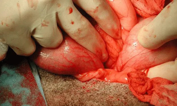
The perinephric mass and the uterine stump were removed via laparotomy (Figures 1E, 1F, and 1G). Histopathology identified a luteoma originating from a right ovarian remnant as well as uterine stump pyometra with endometrial progesterone influence.
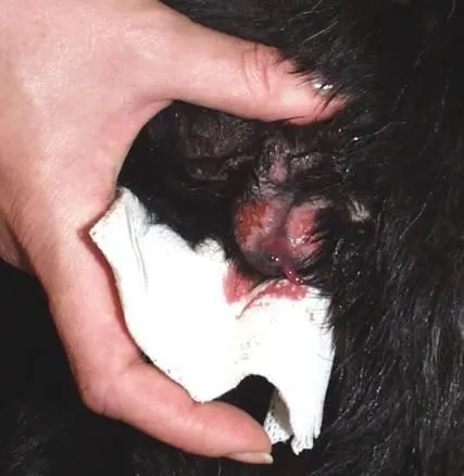
FIGURE 1A
Swollen vulva with serosanguineous discharge present.
Progesterone assay indicated a level of 9.2 ng/mL (normal >2 ng/mL), which is compatible with functional corpora lutea.
Key Point
Ovarian remnants can become functional years after ovariohysterectomy and can also undergo malignant transformation.
Case 2: Foreign Body Bacterial Vaginitis
A 3-year-old Labrador retriever bitch was ovariohysterectomized at 6 months of age. The owners reported that she had been licking her vulva excessively for 3 days and now had pink spotting on her bedding.
Pertinent examination findings included evidence of pain on digital palpation of the vulva and vulvar excoriations. A small amount of purulent discharge was noted (Figure 2A).
Vaginal cytology (Figure 2B) revealed moderate numbers of parabasal cells and numerous neutrophils. Abdominal ultrasonography showed a hyperechoic linear object within the uterine stump, dorsal to the urinary bladder (Figure 2C). Examination findings and vaginal cytology failed to support hormonal influence (no vulvar swelling or cornification).
Vaginoscopy (Figure 2D) exposed a foxtail (grass awn) embedded in the vaginal os of the cervix. It was removed with retrieval forceps under endoscopic guidance.
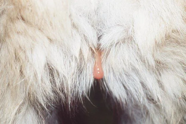
FIGURE 2A
Purulent vulvar discharge; note recessed vulva.
Key Point
Primary bacterial vaginitis in the bitch is rare; an underlying cause is usually present—in this case, a foreign body.
Case 3: Estrogen Exposure
A 5-year-old pug that had been ovariohysterectomized at 7 months of age was presented when the owner noted vulvar enlargement and spotting. In addition, while at the park the pug had become attractive to male dogs during the past 6 weeks.
Pertinent physical findings included vulvar enlargement (Figure 3A) and flagging (receptive behavior). Scant hemorrhagic vulvar discharge was also evident.
Vaginal cytology revealed greater than 90% superficial epithelial cells (Figure 3B), a finding compatible with estrogen influence. The presence of red blood cells and bacteria can be variable during estrus, while the absence of neutrophils suggests the bacteria are opportunistic, not pathogenic.
Abdominal ultrasonography (Figure 3C) showed a prominent but homogenous uterine stump; scant fluid was evident in the uterine stump lumen. There was no evidence of ovarian structures in the region caudolateral to the kidneys.
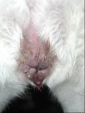
FIGURE 3A
Swollen vulva with sanguineous discharge present.
When specifically questioned, the owner indicated that the housekeeper recently began using transdermal hormone replacement therapy on the forearms and frequently held the pug in her arms. Clinical signs abated over a period of weeks after contact with the dog had been discontinued.
Key Point
Exogenous estrogen exposure can mimic ovarian remnant syndrome in bitches. One clue is the lack of cyclicity, which would be expected with a functional remnant.
Case 4: Uterine Fluid Loss During Pregnancy
A 3-year-old multiparous Cane Corso bitch presented at term gestation after delivering 2 nonviable fetuses. Physical findings included marked abdominal distention, intermittent tenesmus, and malodorous black–green vulvar discharge. No fetus was palpable in the vagina. Brucella canis slide agglutination was negative; subsequent vaginal culture grew Proteus mirabilis organisms.
Cytology of the vulvar discharge (Figure 4A) revealed purulent discharge with numerous toxic neutrophils, intra- and extracellular bacteria, and parabasal vaginal mucosal cells. No cardiac motion was detected on abdominal ultrasonography of 7 nonviable fetuses (Figure 4B). Cardiac motion (Figure 4C) was shown on Doppler imaging of a viable fetus.
Tocodynamometry (uterine monitoring) showed a hypotonic myometrium; no effective uterine contractions were present before or following 10% calcium gluconate and oxytocin administration (Figure 4D).
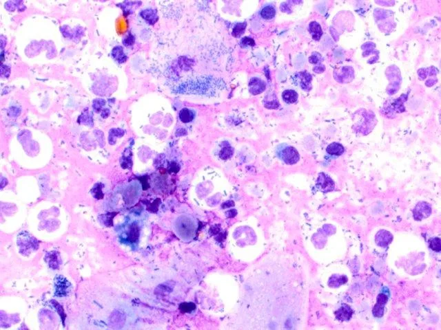
FIGURE 4A
Suppurative, septic inflammation, with numerous neutrophils and intracellular and extracellular bacteria present; vaginal epithelial cells are parabasal.
The nonviable fetuses were removed via hysterotomy. According to the surgeon, less than a normal amount of fluid was present in the uterus surrounding the fetal amniotic sacs. The uterus otherwise appeared normal; it was lavaged and preserved, as this dog was a valuable breeding bitch.
Key Point
Premature cervical relaxation with loss of normal intrauterine fluid and ascending bacterial contamination of the pregnancy was suspected. Uteroverdin (black-green vulvar discharge) is an ominous sign of placental separation.