Vitality of Discolored Teeth in Dogs
Kendall Taney, DVM, DAVDC, FAVD, Center for Veterinary Dentistry & Oral Surgery, Gaithersburg, Maryland
In the Literature
Feigin K, Bell C, Shope B, Henzel S, Snyder C. Analysis and assessment of pulp vitality of 102 intrinsically stained teeth in dogs. J Vet Dent. 2022;39(1):21-33. doi:10.1177/08987564211060387
The Research ...
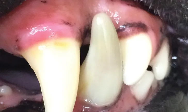
Intrinsically stained teeth are a common finding in dogs and exhibit discoloration within tooth hard tissues, unlike extrinsically stained teeth, which have discoloration on the tooth surface. Causes of intrinsic staining include amelogenesis imperfecta, dentinogenesis imperfecta, tetracycline ingestion during tooth development, dental fluorosis, tooth resorption, hyperbilirubinemia, pulp necrosis, injury, and aging.1
Intrinsic staining can indicate pathology or injury that may affect tooth health or vitality. Determining appropriate treatment can be challenging, as many patients do not have radiologic or clinical signs that can definitively predict tooth vitality.
This prospective study analyzed clinical, radiographic, and histopathologic characteristics of 102 intrinsically stained teeth. There was no evidence of coronal injury (eg, fracture, displacement, mobility) in 55 (53.9%) out of 102 teeth. On histopathologic analysis, 85 (87.6%) of 97 intrinsically stained teeth were nonvital. Radiographic evidence of endodontic disease and periodontal disease was present, respectively, in 58 (57%) and 49 (48%) of 102 intrinsically stained teeth; 29 (28%) had evidence of tooth resorption. Only 19 (18.6%) of 102 intrinsically stained teeth were radiographically normal. All teeth with radiographic evidence of periapical lucency had pulp necrosis. Incisor teeth were most commonly affected, but all tooth types (ie, incisors, canines, premolars, molars) were discolored.
A previous study found that 92.2% of intrinsically discolored teeth were nonvital based on gross appearance of the pulp.2 The current study is the first to histopathologically confirm pulp death, and results verify that a high percentage of intrinsically discolored teeth are nonvital; however, normal contralateral teeth were not histologically evaluated for comparison.
...The Takeaways
Key pearls to put into practice:
Tooth discoloration is a common posttraumatic complication caused by pulpal hemorrhage. Hemolysis of RBCs follows pulpal hemorrhage, and more profound discoloration can occur when hemolyzed RBCs combine with putrefying pulpal tissue.3 Transillumination can help demonstrate increased opacity of an intrinsically stained tooth, but subtle changes can be difficult to detect with the naked eye.
Intrinsically stained teeth are likely nonvital. In this study, most (87.6%) intrinsically stained teeth were histopathologically confirmed to be nonvital, which is consistent with results of a previous study.2
Nonvital teeth are likely to develop endodontic and periodontal disease and should be treated quickly with exodontics or endodontics.
Radiographic signs may support a diagnosis of nonvitality but are not definitive.3,4 All teeth with radiographic evidence of periapical lucency also had pulp necrosis in this study.
You are reading 2-Minute Takeaways, a research summary resource presented by Clinician’s Brief. Clinician’s Brief does not conduct primary research.