Veterinary Dental Teleradiology
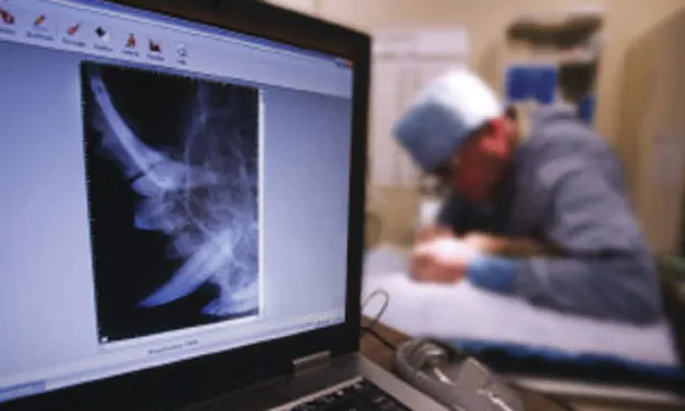
Dental radiology is quickly becoming the standard of practice in veterinary medicine. It has been shown to be 1 of the best ways to improve quality of dental care in veterinary practice. As a result, practices are adding this technology on a daily basis. However, most veterinarians have received minimal to no training in school regarding obtaining and interpreting dental radiographs.
Dental teleradiology is a relatively new but invaluable service available to veterinarians that fills this gap. It is already of great value in human dentistry.1,2 Teleradiology has the potential to improve the quality of patient care and reduce the stress and frustration for practitioners learning to interpret this novel diagnostic format. Teleradiology can also be a profit center for practices that use it.
IndicationsThere are several reasons to use teleradiology, especially if dental radiology is new to your practice.
The most obvious indication is to obtain expert interpretation on unknown, questionable, or novel abnormalities.
Dental specialists can also help practitioners and their staff with the challenges of positioning and exposure.
Finally, veterinary dental teleradiologists can be an invaluable aid when it comes to equipping your dental operatory.
Techniques & EquipmentDental teleradiology can be used by any practitioner with a digital dental radiography system. The efficiency of the teleradiology experience will vary with the system. Some digital systems allow users to directly send images to a teleradiology service by using a DICOM (digital imaging and communication in medicine) protocol. In digital dental systems that support DICOM, sending full DICOM files is recommended.
An alternative method of sending images for review is to export the images as JPEG files and upload them to a teleradiology service. Sending JPEG images may be cumbersome; specialists will not be able to window and level images, and patient demographic data will not be included with the images. Nonetheless, sending JPEG images is a necessity for systems that do not use DICOM.
To get the most out of teleradiology, full-mouth radiographs should be submitted initially as a learning aid.
Advantages_Improved Diagnosis of Abnormalities_The main advantage of dental teleradiology is that specialists often find more abnormalities than most general practitioners. This leads to superior patient care and ultimately should increase income for the practice (because of the increased number of necessary procedures performed).
_Detailed Treatment Recommendations_Furthermore, teleradiology readings deliver an official report for the practitioner and client, with detailed treatment recommendations. Expert advice coming from a third-party specialist should go a long way toward improving client compliance with prescribed treatments. If the teleradiology service offers STAT readings, therapeutic procedures can be performed under the same anesthetic episode.
_Comparison of Radiographs & Photographs_Most teleradiology services allow submission of digital photographs of the oral abnormality along with radiographic images. If the reader is a veterinary dentist, these additional images can help with diagnosis and treatment recommendations. In addition, photos of an oral abnormality with normal radiographic findings can be included for expert opinion.
_Permanent Image Backup_Dental images are as much a part of the medical record as are whole-body films or patient progress notes. An additional advantage of teleradiology services is permanent backup of images. Practices often use laptops or dedicated desktops for dental radiographs. In many practices, these computers are not backed up, and laptops are notoriously unreliable.
Many teleradiology sites permanently back up all submitted images. This can save money and prevent potential liability issues for practices that use these services. Check with the teleradiology service for details on their backup system.
_Official & Legal Interpretation_Finally, teleradiology provides the advantage of reports that can be used as an official, legal interpretation. This can be invaluable if there is ever a dispute about whether a procedure was performed correctly or was necessary. A radiology reading could even be performed after a complaint has occurred, which may quell the client’s concern early on and avoid any disputes over the procedure.
Disadvantages_Lack of Dental Interpretation_Veterinary dental teleradiology has only a few minor disadvantages. The first is that most teleradiology services are staffed by radiologists rather than veterinary dentists. Although radiologists are qualified to read skull images, most are not trained in reading dental radiographs. A significant amount of subtle and unusual abnormalities are found on intraoral films, and dentists are significantly more knowledgeable in this area.

In addition, veterinary dentists will have the most up-to-date treatment recommendations for the various abnormalities. Therefore, it is best to use a service that employs veterinary dentists for reading dental films.
_Time to Export Digital Images_Another disadvantage is the time that it takes to export digital images from various systems. The major reason for this is lack of DICOM connectivity. Export times vary widely; some are fairly quick and others are more laborious. Companies are constantly improving this feature; therefore, any recommendations made in this article would quickly become outdated. It is best to discuss this aspect of the software with the representative of the digital system company before purchasing the product. Uploading to teleradiology sites is very quick once the images are exported.
Economic Impact_Expense_For practices already equipped with digital systems, there is minimal expense. The only cost is technician time to upload the images. With practice, this should take only a few minutes. For practices that do not have a digital system, standard images can be converted into digital images by scanning or taking a high-quality digital photograph of the film.
_Revenue_The typical cost of a reading is $45 to $50, which should be passed on to the client with an additional small fee for technician time. Therefore, these readings become a direct income source for the practice. In addition, most readings lead to an increase in the number of dental procedures performed. Some services will refund the cost of the reading to the client if the reading results in a referral for specialty dental care.
In addition, if the teleradiology site offers permanent backup, it can save the practice information technologies expenses if all films are submitted for review.
ConclusionVeterinary dental teleradiology is the future of veterinary dentistry. Primarily this is because it greatly improves patient care. However, it also helps the staff obtain good-quality radiographs and acts as a permanent, secure backup. Finally, it should generate a significant financial return for the practice. Many readings result in additional procedures performed, which benefits patient health. Perhaps more important, the occasional unnecessary procedure (see The Chevron Effect, below) will be prevented.
FIND MORE
Read Digital Dental Radiology (January 2011) for information about indications, advantages, and technological advances of digital dental radiology. Available at cliniciansbrief.com/journal.
The Chevron Effect
Teleradiology consults for assistance with radiographic interpretation avoid treatment mistakes.
One common misinterpretation of dental radiographs occurs in evaluating the maxillary canines for periapical rarefaction (lucency).3 Periapical rarefaction is typically the sign of endodontic disease in the form of a granuloma (or abscess).
However, radiographs often reveal a small, regular widening of the periodontal space at the apex of the maxillary canines (Figure 1). This common artifact is known as a chevron effect, summation effect, or penumbra. Although this may appear to be a periapical lesion, it is differentiated from a pathologic abnormality because it is regular and v-shaped, as opposed to irregular and round (Figure 2).
Figure 1: The small, regular, v-shaped widening of the periodontal space at the apex of this maxillary canine is an artifact known as the chevron effect (arrows).

Figure 2: True periapical rarefaction; note that the lesion is more rounded and dark (arrows).

This subtle difference can help the clinician choose proper care and avoid undertreating (missing an infection) or overtreating (surgical extraction or root canal therapy of a healthy strategic tooth). One of us has personally spoken to many veterinarians who have extracted these questionable teeth. A teleradiology consult for assistance with radiographic interpretation avoids this confusion and treatment mistake.
The cause of this artifact is unknown; however, a similar effect is seen in the large molar teeth of dogs. In the large molars, the apical region of the vascular meshwork has a vertically longer, more elliptical shape and features adjacent capillary venules with anastomosing branches.4 This is attributed to movement of blood from the periodontal ligament to the alveolar bone, which allows the periodontal ligament to act as a shock absorber of the occlusal forces. Whether this holds true for the canine teeth has not been determined, but it is theoretically sound.
VETERINARY DENTAL TELERADIOLOGY • Matthew Wright & Brook A. Niemiec
References
1. Using teledentistry to improve access to dental care for the underserved. Fricton J, Chen H. Dent Clin North Am 53:537-548, 2009.2. Teledentistry in the United States: A new horizon of dental care. Sanchez Dils E, Lefebvre C, Abeyta K. Intl J Dent Hyg 2:161-164, 2004.
Veterinary dental radiology. Niemiec BA. In Niemec BA (ed): Small Animal Dental, Oral and Maxillofacial Disease: A Color Handbook—London: Manson, 2010, pp 63-87.4. Scanning electron microscopic observation of microvasculature in periodontium. Matsuo M, Takahashi K. Microsc Res Tech 56:3-14, 2002.