Surgical Treatment of Sialoceles: The Ventral Approach
J. Brad Case, DVM, MS, DACVS, University of Florida
Dogs have 4 pairs of major salivary glands (ie, parotid, zygomatic, mandibular, sublingual). The major function of these glands is to lubricate the oral cavity to facilitate digestion and maintain overall health of the mouth. Saliva is predominantly composed of water but also contains mucus and enzymes; when saliva leaks from damaged or diseased ducts, a local inflammatory process begins and creates sialoceles, subcutaneous collections of saliva lined by thin-walled sacs of granulation tissue.
Sialoceles
In dogs, sialoceles occur most commonly in the cervical and/or sublingual regions but can also occur in the pharyngeal and zygomatic regions.1 Clinical signs associated with sialoceles include swelling, dysphagia, oral hemorrhage, and, in patients with pharyngeal sialoceles (see Pharyngeal Sialoceles), respiratory distress. Exophthalmos can be seen with zygomatic sialoceles. Although the exact cause of sialoceles is often unknown, a number of causes and associated factors exist and include trauma, foreign body, neoplasia, idiopathic causes, and sialolithiasis. Male dogs and standard poodles, German shepherd dogs, and dachshunds appear to be predisposed.1
Pharyngeal Sialoceles
Pharyngeal sialoceles are diagnosed through oropharyngeal examination and identification of lateral pharyngeal swelling containing salivary fluid. Although rare, significant pharyngeal swelling can occur with disease and leakage of the sublingual salivary glands and may necessitate treatment. Drainage and marsupialization of sialoceles in the oropharynx is the most common treatment performed and can be combined with sialoadenectomy. Drainage and marsupialization can be effective, but if recurrence is observed, surgical removal of the affected mandibular and sublingual glands is indicated.
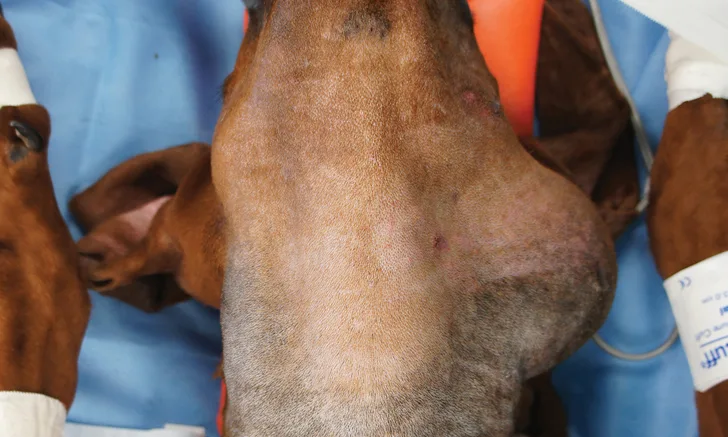
Determining the location of the swelling is an important part of localizing the sialocele to the affected gland(s). For example, cervical and sublingual sialoceles (ie, ranulas [see Ranulas]) are the result of rupture of one or both of the sublingual salivary glands. The sublingual salivary glands are the most commonly affected glands in most (>90%) sialocele cases in dogs.1 Cervical swelling can often be localized to one side by placing the dog in dorsal recumbency and allowing gravity to move the swelling to the affected side (Figure). In the case of ranulas, the affected side should be obvious on oral examination. Determining the side of origin may be more difficult in bilaterally affected dogs or those with large sialoceles.
Ranulas
Ranulas are diagnosed through visual observation of mucosal swelling ventral to the tongue base and adjacent to the frenulum. Ranulas typically develop secondary to injury of the polystomatic portions of the sublingual gland, which results in submucosal accumulation of saliva. Oral marsupialization can be attempted alone or in conjunction with sialoadenectomy. Ranulas should be incised and saucerized to expose the mucocele lining. The cut edges of the mucocele should be sutured to the sublingual mucosa, creating a stoma. Marsupialization is rarely curative as a sole treatment, and if recurrence is observed, surgical removal of the affected mandibular and sublingual glands is indicated.

Dog with a left-sided cervical sialocele. Gravity-dependent swelling can be noted on the affected side.
Diagnostic investigation should include a thorough oral examination, cervical and thoracic radiography or CT, CBC, serum chemistry profile, urinalysis, and fine-needle aspiration/cytology of the swelling. Sialoceles can often be diagnosed through visualization of a viscous, brown to blood-tinged mucoid fluid following fine-needle aspiration. Periodic acid–Schiff staining may be needed in some cases for definitive diagnosis.
Most dogs with sialoceles are otherwise healthy and surgical excision can be performed following diagnostic investigation. Although simple drainage has been reported to be effective, it is associated with a high recurrence rate and should be avoided. Salivary gland removal should be performed before surgical drainage, as significant scar tissue and adhesions can form during surgical drainage due to irritation of adjacent tissue by the inflammatory saliva. Consequently, complete removal of the affected glandular tissue during the initial surgery is critical. Dogs with large sialoceles and those that are bilaterally affected may require excision of both the right and left sublingual and mandibular salivary glands. Although the mandibular glands are typically not involved with sialoceles, their anatomic association with the affected sublingual gland(s) makes removal of both the mandibular and sublingual glands necessary. Dogs can undergo excision of both sublingual and mandibular glands without experiencing xerostomia, provided the parotid and zygomatic glands are left intact.
The dissection of sialoceles can be challenging, as important neurovascular anatomy surrounds the salivary glands; for example, the mandibular lymph node is located immediately craniad and ventral to the sublingual and mandibular glands and could be mistaken for the salivary gland by an inexperienced clinician. Thus, it is critically important that the surgeon have a complete understanding of the relevant anatomy before considering sialoadenectomy in a clinically affected dog. Prognosis following sialocele excision, however, is excellent in most cases, with the risk for recurrence being <5% for an experienced surgeon. Most dogs are stable and systemically healthy postoperatively, so fluid support beyond the time of voluntary eating is unnecessary. Intravenous opioid analgesics are rarely indicated beyond 24 to 48 hours after surgery. Nonsteroidal therapy for 3 to 5 days is typically adequate, provided there are no contraindications for their use.
Surgical Approaches
Two surgical approaches—lateral approach and ventral mandibular approach—have been described for mandibular and sublingual gland excision. The lateral approach offers simpler dissection, whereas the ventral approach offers greater and simultaneous exposure of both the right and left mandibular and sublingual glands. Although either approach is acceptable if care is taken to ensure complete removal of the sublingual gland(s), extensive craniad dissection may be required in some cases; thus, the ventral approach is preferred by the author and described in this article.
Step-by-Step: Ventral Approach to Sialodaenectomy
What You Will Need
Standard general surgery pack with needle holders, DeBakey thumb forceps, Metzenbaum scissors, suture scissors, surgical blade, and hemostatic forceps
Medium self-retaining retractors (eg, Gelpi, Weitlaner) and electrosurgical pencil
Sterile gauze, cotton tip applicators, and surgical suction
Step 1
Place the patient in dorsal recumbency. This will cause the swelling to fall to the affected side unless bilateral or significantly swollen cervical sialoceles are present, in which case swelling may be present on the midline.
If removing both the left and right glands, make the incision on the midline from the midmandible and extend it caudally 1 to 2 cm caudal to the palpable mandibular salivary glands. If removing only the left or right glands, direct the incision slightly to the affected side while extending it caudally.
Step 2
After incising the skin and subcutaneous tissue, palpate the affected gland. Palpation is generally easy but may be difficult in some cases, as the affected gland may be displaced dorsally along the patient’s neck due to dorsal recumbent positioning. To identify the affected sublingual/mandibular gland(s), use digital pressure along the patient’s neck to displace the gland ventrally and allow for easier palpation in the incision.

Step 3
Continue the approach carefully using a combination of sharp and blunt dissection through the mandibular/sublingual gland capsule until the glandular tissue is exposed. Apply gentle caudal traction to the mandibular gland with a stay suture or Allis tissue forceps, and continue the dissection cranially to the level of the digastricus muscle. The smaller and more friable sublingual gland is located at the cranial end of the mandibular gland; avoid direct traction on sublingual gland tissue.

Author Insights
It is important to isolate and dissect along the path of the mandibular/sublingual duct and to not use sharp dissection in an orientation other than parallel to the duct. Inadvertent transection of the duct/gland complex can cause incomplete removal and/or recurrence of the sialocele. The risk for incomplete removal with an experienced surgeon is low.
Incision of the sialocele is required during dissection of the gland/duct. It is not necessary to excise the distended granulation tissue composing the sialocele, but incision of the tissue is required to isolate the affected region of the salivary gland. Suction of the mucoid salivary fluid can be accomplished by sterile in-house suction and a Frazier tip.
Step 4
Identify the caudolateral aspect of the digastricus muscle and isolate the duct to this level. Carefully isolate the mylohyoideus muscle—a thin muscle with fibers that run in a transverse orientation—with blunt dissection before incising it to avoid injury to underlying structures (eg, hypoglossal nerve, lingual nerve and artery). Then, incise the mylohyoideus muscle cranially to the midintermandibular region.
Step 5
Insert hemostatic forceps from cranial to caudal under the digastricus muscle to grasp the gland/duct complex (A). Exercise caution to limit connective tissue being entrapped in the jaws of the forceps. Amputate the gland/duct caudal to the hemostatic forceps, and use the forceps to retract the gland/duct craniomedially under the digastricus muscle. Identify the lingual nerve, and dissect and ligate the duct at the level of this nerve (B). Excise the remaining sublingual salivary tissue/duct and submit for pathology.


Step 6
Evaluate the surgical site for bleeding and address as needed. Use sterile saline to lavage the wound, and reappose the mylohyoideus muscle with a fine monofilament absorbable suture. Close the subcutaneous tissue in a simple continuous fashion with a fine monofilament suture, and close the skin with an intradermal or external pattern.

Step 7
Continue intravenous fluid administration postoperatively and adjust to meet the demands of the patient.
Editor's note: This article was originally published in September 2019 as "Ventral Approach to Sialoadenectomy."