Using a Wood’s Lamp
Karen A. Moriello, DVM, DACVD, University of Wisconsin–Madison
A Wood’s lamp is a hand-held device that emits long-wave ultraviolet radiation through a nickel or cobalt glass filter. The filter is opaque to all light except a band between 320 and 400 nm. Fluorescence occurs when shorter wavelengths are absorbed and radiation of longer wavelengths (visible light) is emitted.
The Wood’s lamp was invented in 1903 by Baltimore physicist Robert Wood. In human medicine it is used to detect dermatophyte infections, bacterial infections, porphyria, and pigmentary diseases. In veterinary medicine, Wood’s lamps are most commonly used to help identify dermatophytosis caused by Microsporum canis. (Trichophyton species and M gypseum infections do not produce metabolites that cause fluorescence.) They are also useful when examining hairs under the microscope for evidence of ectothrix infections and occasionally as an aid in monitoring therapy.
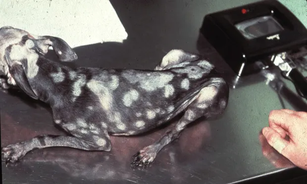
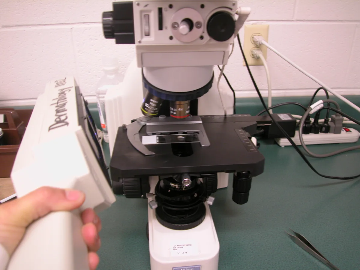
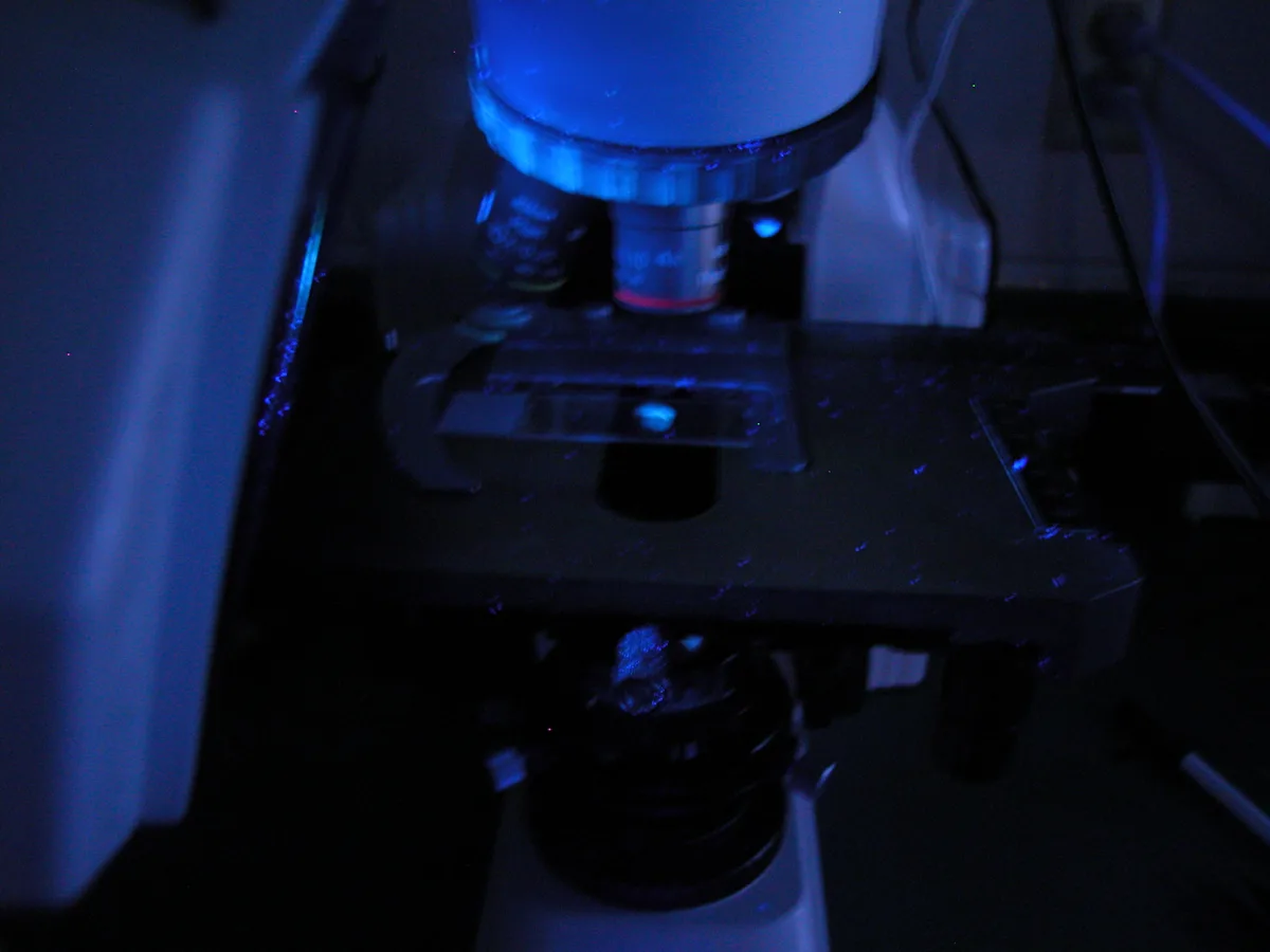
I recommend using a good quality electric Wood’s lamp because of inconsistent results with battery-operated models. There are 2 types of hand-held lamps available—one has only a lamp (Figure A) and the other has a light surrounding the magnifying lens (Figure B). Both are acceptable; however, care must be taken to clean the magnifying lens in the latter type, which is also larger and sometimes more difficult to use.


Wood’s lamps produce a wide range of fluorescent colors depending upon the substrate being examined. Many things fluoresce under a Wood’s lamp including scales, sebum, seborrhea, crusts, and topical agents. True M canis fluorescence is bright apple green and infected hairs glow from the bulb to the tip. Skin oils often produce a blue-green to orange fluorescence. Some fabric fibers, especially synthetic carpets, produce bright apple-green fluorescence, the color most consistently associated with M canis infections. A Wood’s lamp is only a screening tool for M canis infections, however, because not all strains will fluoresce.
Points to Remember
Wood’s lamp examinations are effective only as a screening tool for M canis infections. Negative fluorescence does not rule out dermatophytosis.
Wood’s lamp examinations are cost- and time-effective when examining known culture-positive animals (M canis) or highly suspect animals (eg, cats with skin lesions).
Wood’s lamps can be used in any species susceptible to M canis infections, but because dermatophytosis is more common in cats and more pleomorphic in presentation, it is considered a core diagnostic test in cats with skin disease.
Fungal culture is always recommended to confirm the infection.
Allow the lamp to warm up for 3 to 5 minutes to be sure it is being used at full power. Perform the test in complete darkness to allow for the examiner’s retinas to be totally dark-adapted. The gradual “development” of fluorescence is the result of one’s eyes becoming adapted to the light.
Most mistakes made with the Wood’s lamp are due to failure to allow the lamp to warm up completely, not having the room dark enough, not taking enough time to perform a complete examination, and not allowing time for the examiner’s eyes to adapt to darkness.
Step-by-Step: Wood's Lamp as a Screening Tool
Step 1
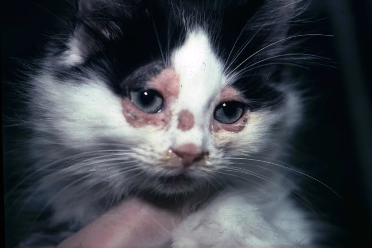
Under proper conditions, examine known culture-positive animals to help locate lesions or use as a screening tool in a cat with skin disease. Examine clinically lesional areas. Particularly in kittens, be sure to examine the hairs inside the “bell” of the ear and between the toes. Early lesions of dermatophytosis are often found on hairs on the face, especially around the eyes, in and near the ears, and around the lips.
Step 2
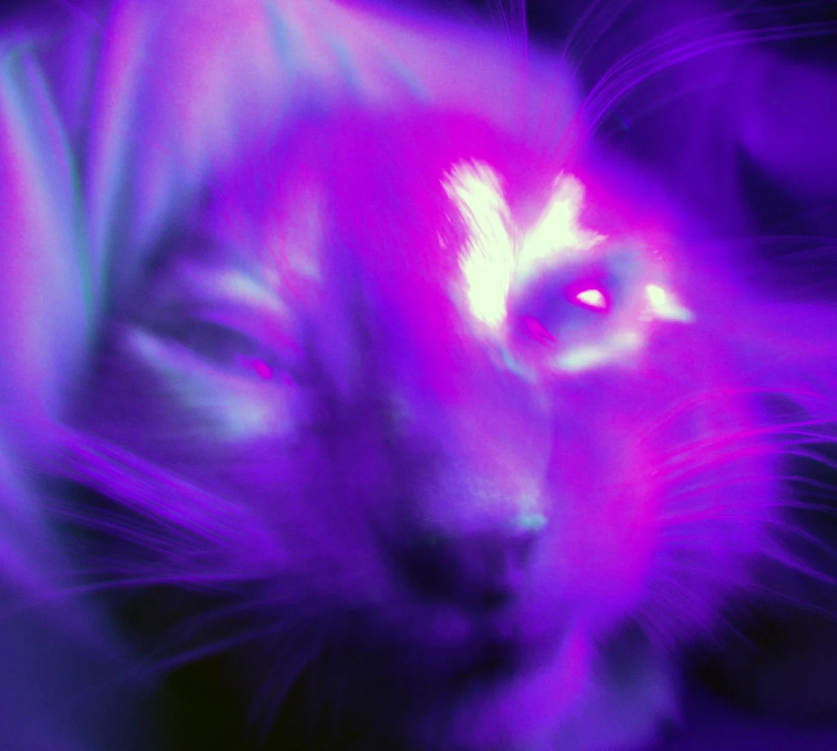
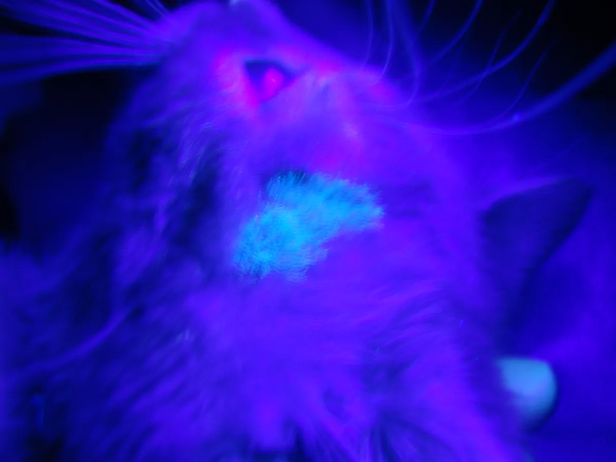
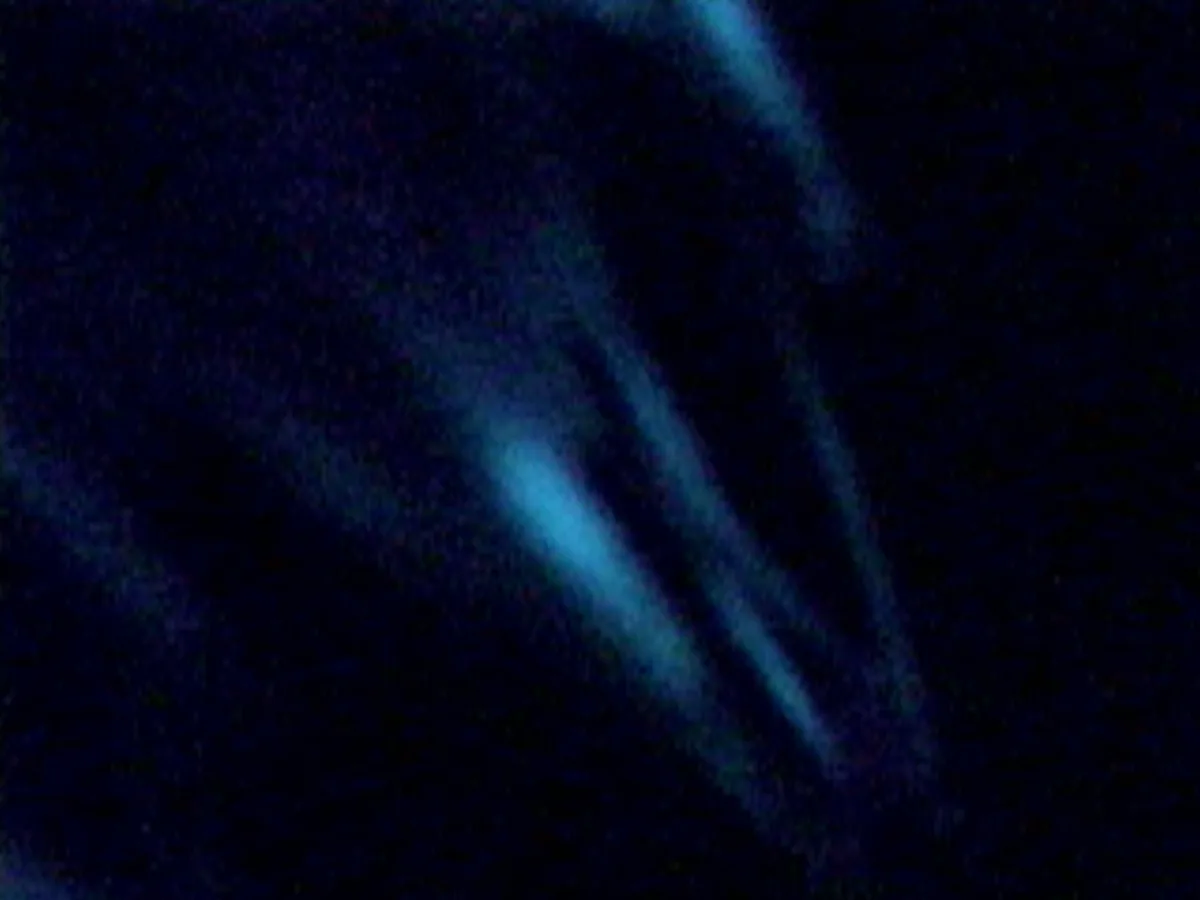
True fluorescence of M canis is apple green in color, as seen in the glowing hairs of an infected cat. Look under crusts for glowing hairs.
Procedure Pearl
Good restraint is necessary for a careful examination. You will need one person to hold the animal, one to hold the lamp, and one to pluck suspect hairs.

Step 3
Topical forms of common medications such as doxycycline (forelimbs of the cat, A) and tetracycline (over the left eye, B), can give “false fluorescence.” Get a good medication history.
Step 4
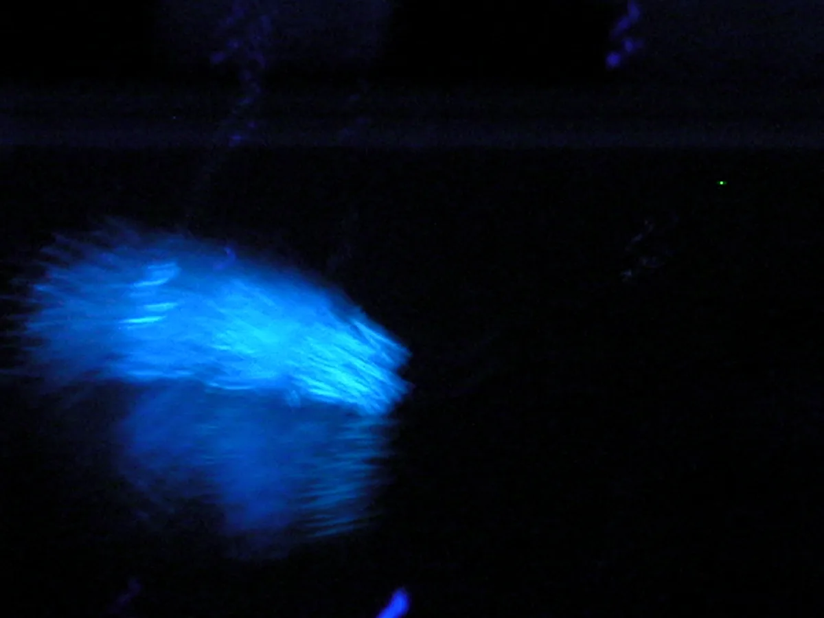
A mat of glowing hairs is depicted in the photomicrograph. Glowing hairs can be plucked for culture or for direct examination for spores and fungal hyphae.
Step-by-Step: Wood’s Lamp to Directly Examine Hairs
Step 1
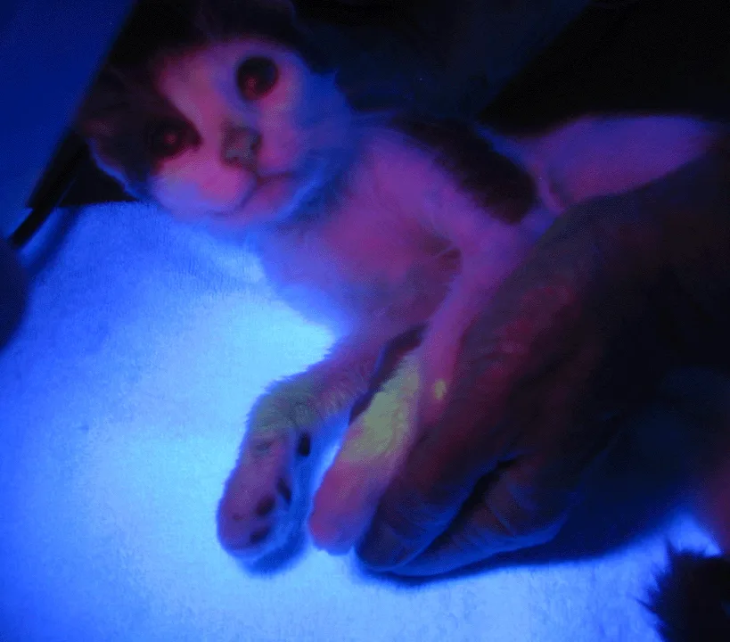
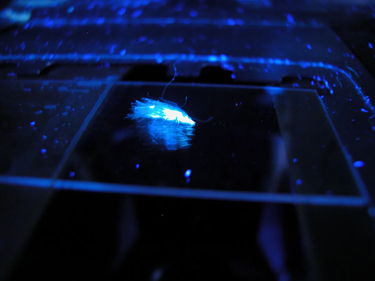
Pluck hairs in the direction of growth to see if the hair bulb is glowing. Try to pluck individual hairs as opposed to large mats of hairs (A). Move the lamp to the area where the microscope is located and turn out the lights. Hold the Wood’s lamp over the slide, allowing you to locate the hairs to examine (B).
Step 2
Place the slide with the glowing hair on the microscope stage. If the hair cannot be found easily, turn the microscope light on and the laboratory lights off. From the side or from beneath, shine the Wood’s lamp on the slide while looking through the scope (A). Note the glowing hairs on the microscope slide (B).
Step 3
While looking down the microscope column, move the slide on the stage until a fluorescent object is seen. This is best done at 10× to avoid damage to the microscope lens.
Step 4
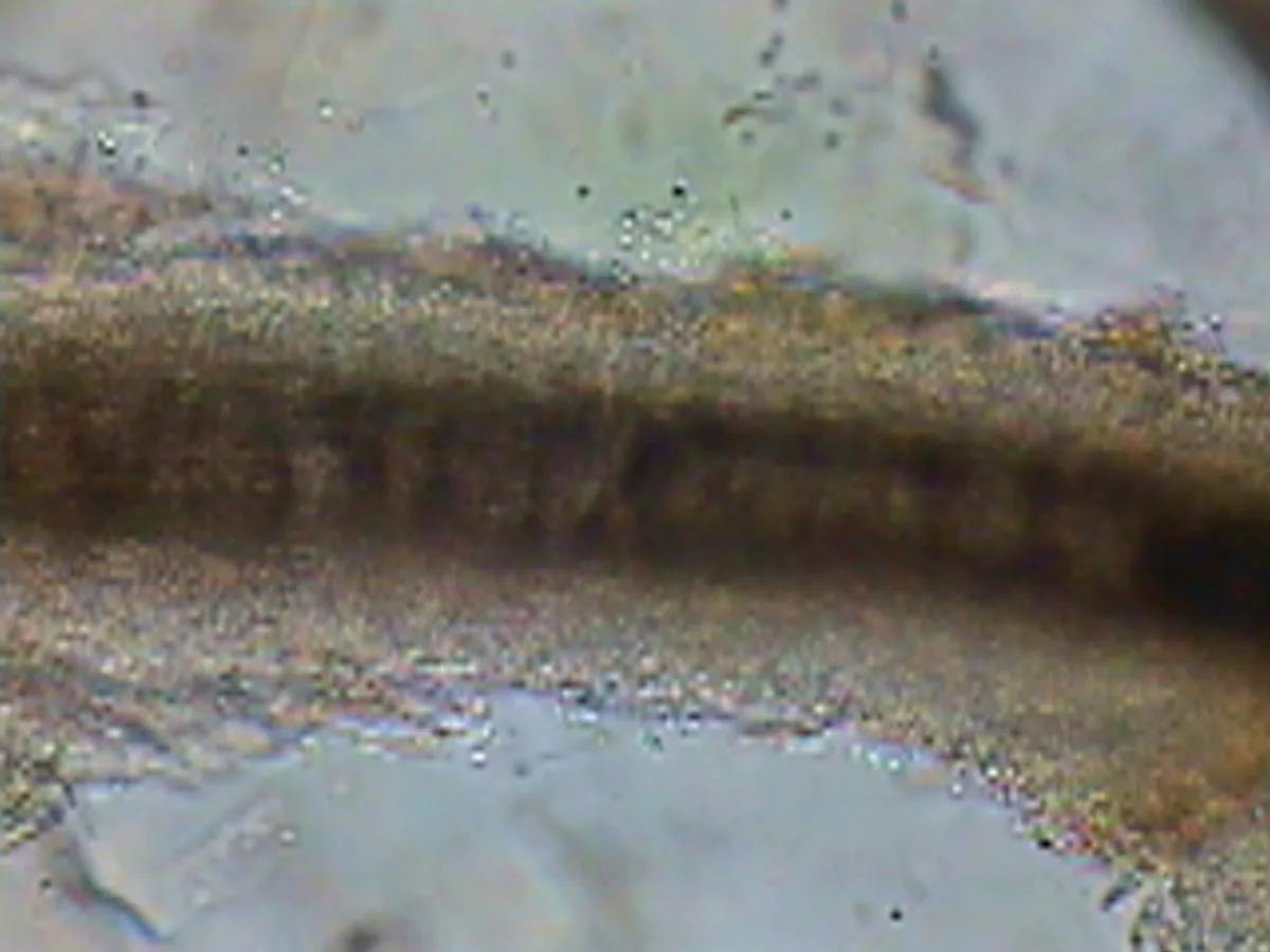
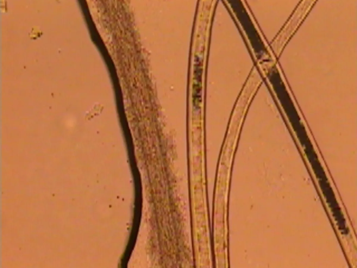
Turn the lights off in the laboratory and the microscope light on and examine “glowing hair.” The micrographs depict a cuff of ectothrix spores from M canis (A), and 3 normal and 1 infected hairs (B). Note that the infected hair is wider and more “filamentous” in appearance than the normal one.
Procedure Pearl
Confirming evidence of infection through direct examination with a Wood’s lamp allows treatment to begin before results of the fungal culture are available.
Use to Directly Examine Hairs
Hairs plucked for direct examination can be examined in mineral oil or in a clearing agent, such as potassium hydroxide or chlorphenolac (requires formulation by compounding pharmacy).
Clearing agents aid in examination of infected hairs by making the background debris appear homogenous and infected hairs appear more refractile. Clearing agents can be caustic and can damage the microscope lens if not used properly, but infected hairs can be examined with mineral oil if mounted in a small amount of oil.
The Wood’s lamp can then be used to help find the glowing hair(s) on the slide and/or through the microscope lens. (Wood’s lamps are equally helpful with clearing agents.)
M canis produces “cuffs” of spores on the outside of the infected hair, indicating ectothrix invasion. As the infected hair is damaged, it will appear as if the entire structure is replaced by spores and hyphae.
If an infection is confirmed, start therapy prior to final fungal culture results.
Use to Monitor Treatment
In animals with a fluorescing strain of M canis, a Wood’s lamp can be used in addition to fungal cultures to monitor treatment. This requires careful examination of the animal prior to treatment and documentation of lesions and location. Early in the course of an infection, the entire hair will glow, including the hair bulb. As the infection resolves, progressively fewer Wood’s-positive hairs are visible and fluorescence is seen on just the distal shafts and eventually on only the tips.
If an animal is clinically cured but still fungal-culture–positive, a Wood’s lamp examination can help identify whether there is a nidus of infected hairs. In these situations, infected hairs are often found near the eyes and ears; owners often inadequately treat these areas topically. Note the presence of lesions on the faces of the kittens shown. Animals that appear to be clinically cured but are persistently culture-positive often have small lesions around their faces and ears due to lack of thorough application of topical therapy.