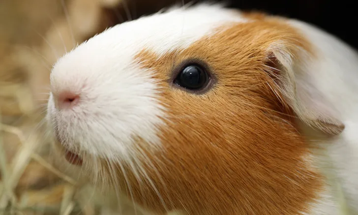
Urolithiasis in Guinea Pigs
Bruce H. Williams, DVM, DACVP, Joint Pathology Center, Rockville, Maryland

In the Literature
Rooney TA, Eshar D, Wong AD, Gardhouse S, Beaufrère H. The association between bloodwork, signalment, and urolithiasis in guinea pigs (Cavia porcellus). J Exotic Pet Med. 2021;38:26-31.
The Research …
Urolithiasis is a common disease in guinea pigs, representing up to 4.73% of all diagnoses in this species.1 Not much is known about this condition in guinea pigs, but many potential causes have been suggested, although not proven, including a diet heavy in leafy greens or alfalfa, overconditioning, poor sanitation, stationary lifestyle, urine retention, adrenal disease, and dehydration.
This retrospective study sought to determine whether signalment or blood work analytes were associated with evidence of urolithiasis identified via diagnostic imaging. Of the 81 guinea pigs included, 32 (40%) had evidence of urolithiasis on imaging. The likelihood of urolithiasis increased when packed cell volume decreased, plasma creatinine concentration increased, and plasma phosphorus concentration decreased. Despite this statistical significance, the predictive value for discriminating between guinea pigs with and without urolithiasis was low.
… The Takeaways
Key pearls to put into practice:
Urolithiasis is common in guinea pigs of both sexes, but females are thought to be more predisposed. In this study, however, affected males outnumbered affected females by almost a 2:1 margin.
Although uroliths in guinea pigs have historically been reported to be composed of calcium oxalate, this study found uroliths to be almost exclusively composed of calcium carbonate.
Decreased packed cell volume and serum phosphorus, along with increased serum creatinine, is suggestive of urolithiasis; imaging is strongly recommended to confirm diagnosis.
You are reading 2-Minute Takeaways, a research summary resource presented by Clinician’s Brief. Clinician’s Brief does not conduct primary research.