Urethral Obstruction in Cats
Catherine V. Sabino, DVM, DACVECC, Red Bank Veterinary Hospital, Tinton Falls, New Jersey
Urinary obstruction is a life-threatening emergency. The inability to empty the bladder results in increased pressure in the urinary tract and a decreased glomerular filtration rate, which causes a buildup of toxins in the blood. The electrolyte abnormalities and fluid imbalances that result because the patient cannot eliminate the toxic substances may lead to hypotension, acute kidney failure, cardiac arrhythmias, and death.1 Urinary obstruction can occur in cats of any age but is most common in male cats 2 to 10 years of age.2
Signs & Causes
The most easily recognized signs of urinary obstruction are vocalization and straining while posturing to urinate. Cats may frequently be in and out of the litter box and may attempt to void outside the litter box. A patient straining without producing urine requires immediate medical attention; therefore, the reason for straining must be differentiated from other causes (eg, constipation, diarrhea, urinary tract discomfort associated with cystitis or a urinary tract infection) that also require medical attention but are not immediately life-threatening. Other signs include vomiting, lethargy, anorexia, and, in more advanced cases, weakness or collapse.3,4

Microscopic image showing struvite crystals found in the urine of a cat with lower urinary tract signs. Photo courtesy of Catherine V. Sabino, DVM, DACVECC
Physical causes of obstruction include urethral plugs, bladder stones/uroliths (see Figure 1) lodged in the urethra, strictures (ie, scarring) of the urinary tract secondary to previous infection or trauma, or, less frequently, cancer.2,5
Obstructions may also result from a functional disorder of the urethra (eg, muscle spasms, swelling), which blocks the flow of urine. In patients with a functional obstruction, no physical blockage is present.
In many cases, the underlying cause cannot be identified and may be attributed to feline interstitial cystitis (FIC), a sterile inflammation of the bladder whose cause remains unknown. Excessive body weight, low activity levels, stressful situations (eg, environmental changes, conflicts between cats in multicat households), decreased water intake, and consumption of a primarily dry-food diet are thought to predispose patients to FIC or functional obstruction, although not all studies agree.3,6-8
Physical obstructions can be partial or complete and taking care to differentiate is vital.
Diagnosis
Diagnosis is based on a nonexpressible urinary bladder, which typically is large and firm. Clinical signs may include:
Dull/depressed mentation
Dehydration
Bradycardia (ie, heart rate <140 beats per minute)*
Cardiac arrhythmias*
Hypothermia (ie, <96.6<sup⁰sup>F [35.9<sup⁰sup>C])*
Increased respiratory rate (ie, >40 breaths per minute)*
Pale mucous membranes and prolonged capillary refill time
Weak pulses*
Diagnostic testing helps identify the extent of abnormalities caused by the obstruction. At a minimum, serum electrolyte concentrations should be measured for evaluation of life-threatening hyperkalemia. Evaluation of packed cell volume, total solids, and kidney values will allow further characterization of the severity of the patient’s condition. CBC, serum chemistry profile, and blood gas analysis may also be performed to fully evaluate the patient. Potential abnormalities include:
Hyperkalemia (elevated potassium): Decreased excretion through the kidneys, reabsorption of potassium from the bladder, and decreased blood pH result in elevated potassium. Blood potassium elevation causes muscle weakness and cardiac conduction abnormalities that can be seen on an ECG.10
Metabolic Acidosis (decreased blood pH): Obstruction causes decreased excretion of hydrogen ions through the kidneys. The systemic consequences of severe metabolic acidosis are decreased blood pressure, cardiac output, and hepatic and renal blood flow,11 which will increase lactate production and further contribute to acidosis.
Azotemia & Hyperphosphatemia: Urea, creatinine, and phosphorus can increase because of dehydration (prerenal azotemia) and accumulate because of impaired excretion (postrenal azotemia) and development of decreased renal function (renal azotemia).12
Decreased Ionized Calcium: Ionized calcium is necessary for neurologic, muscular, and cardiovascular function. Low levels of calcium concentrations may cause decreased cardiac output, muscle weakness, and tremors or seizures.13
Hyperlactatemia: Reduced tissue perfusion secondary to shock or decreased cardiac output, with impaired excretion ability, can lead to increased blood lactate.11
An ECG should be obtained before initiating treatment. The ECG may show changes in hyperkalemic patients, starting with a narrowing (ie, tenting) of the T-waves, and progressing to a prolonged PR interval, widening of the QRS complex (caused by slowed conduction through the heart), and a decrease in P-wave amplitude. The P-waves may disappear in patients with severe hyperkalemia when atrial conduction ceases. If hyperkalemia is allowed to progress, the QRS complex may widen further and progress to ventricular asystole or fibrillation. The ECG can be used to visualize the most life-threatening consequences of hyperkalemia10; however, the ECG of hyperkalemic patients with urethral obstruction does not display these changes consistently,14 and laboratory tests are necessary to confirm the presence of hyperkalemia.
Treatment
Treatment is based on the underlying cause. All patients should have an IV catheter placed and receive fluid therapy and pain medication. Patients with a partial obstruction may require only pain medication and medications to decrease urethral tone (ie, α-1 adrenergic antagonists [eg, phenoxybenzamine, prazosin]) or to relax the urethral sphincter (eg, acepromazine). These patients should be closely monitored to ensure they are able to urinate before being discharged.
Patients with a complete obstruction, regardless of cause, require placement of a urinary catheter to clear the obstruction and allow the bladder to empty. In the short-term, cystocentesis may be used to empty the bladder if urethral catheterization is not possible.15 Further medical and/or surgical intervention should be provided as needed.
Many patients are hospitalized during treatment. The decision to admit a patient and the length of stay are based on physical examination findings and diagnostic test results. A urinary catheter may remain in place during hospitalization while fluids, pain medications, muscle relaxants, and anti-inflammatories, if necessary, are administered. Patients with severe metabolic derangements will require IV fluids for correction of dehydration and electrolyte abnormalities. Patients with severe hyperkalemia and acidemia may require further treatments (eg, dextrose, insulin, calcium gluconate, sodium bicarbonate) to correct life-threatening abnormalities.

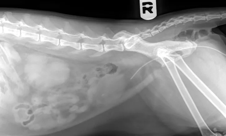
Radiograph showing bladder stones composed of struvite (magnesium ammonium phosphate). Radiograph courtesy of Red Bank Veterinary Hospital, Tinton Falls, New Jersey
Once the obstruction has been cleared, additional diagnostic tests may be performed (eg, urinalysis, urine culture, abdominal radiography and/or ultrasonography) to investigate underlying conditions and identify bladder stones and neoplasms. (See Figure 2.) In patients requiring urinary surgery, the urinary catheter is often left in place until the procedure has been performed.
Prognosis & Long-Term Management
With treatment, more than 90% of patients with urinary obstruction recover normal function3,4; however, the recurrence rate is ≈15% to 35%.16,17 Recurrence of bladder stones can cause physical reobstruction. FIC patients may experience recurrent functional obstruction because of difficulty balancing the many factors that contribute to FIC or because other factors may not be readily identified.
Changes in a patient’s lifestyle may help minimize the risk for recurrence of functional or physical obstruction. For example, diets formulated to reduce urolithiasis may be appropriate for patients with a history of physical obstruction. Management of the indoor cat environment (eg, number of litter boxes; type of litter; scratching, resting, and play opportunities; cat interactions) may reduce stressors and help decrease risk for FIC development, which can contribute to functional obstruction or recurrence. (See Resource.)
Clients should be advised to monitor their pets for signs of a recurring obstruction and seek prompt medical treatment.
Conclusion
In patients with urinary obstruction, toxins accumulate in the bloodstream and, if left untreated, can result in death. Clients may have difficulty differentiating between straining to defecate and straining to urinate with or without urinary obstruction and should be advised that prompt medical intervention may lessen the likelihood of fatal complications, and definitive treatment will depend on the underlying cause.
*May be indicative of severe hyperkalemia9