Ulcerated Dermal Nodule
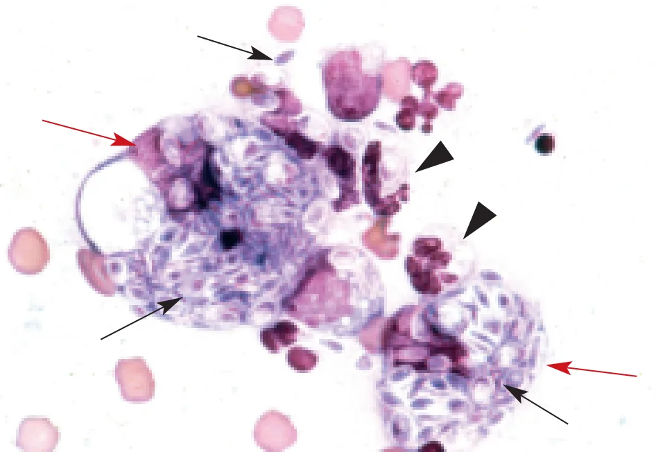
Figure 1
History
Sunshine has no medical problems except for a food allergy, which is treated with monthly steroid injections and a hypoallergenic diet. The cat lives both indoors and outdoors. According to the owner, the lesion has been present for 1 month; it has increased in size since that time and was initially not ulcerated.
Physical Examination
The presenting skin mass is a dermal nodule at the base of the tail, approximately 2.5 cm in diameter. It has an ulcerated surface; a serosanguineous exudate is also present. The lesion is not painful upon palpation. The veterinarian aspirates the lesion with a 22-gauge needle and submits the sample to the local veterinary clinical pathologist for evaluation. The veterinarian looks at the slide. Inflammation may be present, but he decides to wait to treat the patient on the basis of the pathologist's report. He decides to schedule a recheck appointment for the next day and sends the cat home with antibiotics. Figure 1 shows what the pathologist saw on her microscope.

Figure 1
ASK YOURSELF ...
Is the lesion inflammatory or neoplastic?
Describe the structures within the macrophages.
What is the diagnosis?
What other organism could appear similar cytologically?
Diagnosis: Sporotrichosis
The Disease
Sporotrichosis is a zoonotic, dimorphic fungus caused by the organism Sporothrix schenckii. It has been reported in dogs, cats, horses, mules, cattle, buffalo, and humans. The organism is found in decaying plant material and soil. There are three clinical forms: cutaneous, cutaneolymphatic, and disseminated. Cutaneous infection and the cutaneolymphatic form occur via puncture wound and contact with plant and soil matter; the disseminated form occurs via inhalation of organism-laden dust. The infection is more common in immunocompromised animals, except for cats, which can contract the disease even with an intact immune system.
In humans, immunosuppression and occupation (rose gardener, plant nursery worker, horticulturist, farmer) are predisposing factors. The organisms incite a pyogranulomatous response and lesions may mimic draining abscesses, thereby stressing the importance of cytologic evaluation for all lesions presented to the veterinarian. A definitive diagnosis is also important in light of the potential for zoonotic disease.
The Organism
Organisms are most numerous in cats and can be both intracellular (within the macrophages) and extracellular. The organisms appear as pleomorphic yeast, ranging from 3 to 5 µm in diameter. The size and fusiform shape are clues to diagnosis, although round/oval forms may also be seen (Figure 1; black arrows). In other species, only a few organisms may be observed and fungal culture may be necessary for a definitive diagnosis. Figure 2 shows a macrophage infected with a single yeast. Samples may be obtained via fine-needle aspiration biopsy or impression smears/swab smears if a draining tract is present.
The lesion on this cat was excised, and the cat was given supersaturated potassium iodide at 20 mg/kg PO Q 12 H for 30 days. Treatment was curative.
Zoonotic Potential
Infected cats have been reported to be a source of infection for humans. This is because in cats the lesions are associated with high numbers of organisms, in contrast to other species in which far fewer organisms are found. Therefore, all persons in contact with the cat during treatment are advised to wear surgical gloves and wash their forearms with an antiseptic after handling the cat. Infection can occur through direct contact and does not require a preexisting wound.
Did you answer. . .
The lesion appears inflammatory. It comprises an admixture of macrophages (red arrows) and neutrophils (arrowheads). A small amount of peripheral blood is present in the background. A neoplastic population would appear monomorphic.
The structures are yeast organisms that may be round, oval, or fusiform (cigar-shaped; black arrows). They are approximately 3 to 5 µm in diameter and have a slightly eccentrically placed, purple nucleus; pale-blue cytoplasm; and a thin, clear cell wall.
Sporothrix schenckii. The fusiform shape of the yeast is classic for this diagnosis.
These organisms may be confused with Histoplasma capsulatum, especially if only a few round (i.e., not fusiform) organisms are present. Histoplasma organisms are round, 2 to 4 µm in diameter; have an eccentrically placed purple nucleus; have a thin, refractile, clear cell wall; and are typically abundant.
SPOROTRICHOSIS • Kimberly J. Caruso