Treating Feline Triaditis
Harry Cridge, MVB, MS, PG Cert Vet Ed, DACVIM (SAIM), DECVIM-CA, FHEA, MRCVS, Michigan State University
Case
An 8-year-old, 10.7-lb (4.85-kg), indoor-only, neutered male domestic shorthair cat is presented for progressive lethargy and inappetence. Intermittent vomiting and loose stools have occurred for several months, and the cat has lost 2 lb (0.91 kg).
Physical examination reveals normal vital parameters, ≈5% dehydration, icterus, and cranial abdominal discomfort. A minimum database (ie, CBC, serum chemistry profile, urinalysis) and retrovirus screening are performed.
Diagnostics
CBC results show a mild, nonregenerative anemia, a normal platelet count, and a marked inflammatory leukogram with a left shift (Table 1). Blood smear results reveal toxic neutrophils. Lymphocyte and eosinophil concentrations are within normal limits.
Serum chemistry results show elevated ALT, ALP, and gamma-glutamyl transferase (Table 2). Total serum bilirubin and serum creatinine are elevated, with a concurrent urine specific gravity of 1.042. All other variables are within normal limits, including creatine kinase activity. A point-of-care test is negative for FeLV antigen (p27) and FIV antibodies.
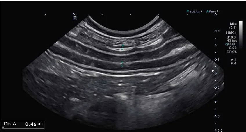
Moderate hepatomegaly is seen on abdominal radiographs, and there is no evidence of mechanical obstruction. Abdominal ultrasound (Figure 1) reveals an enlarged, hypoechoic pancreas with a hyperechoic surrounding mesentery. The gallbladder is bilobed and contains a moderate amount of non–gravity-dependent hyperechoic luminal debris, and the wall is mildly thickened. Intestinal wall thickness is also increased, with a prominent muscularis layer.
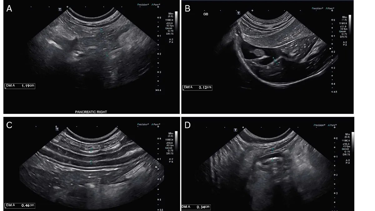
FIGURE 1 Ultrasonographic findings showing an enlarged hypoechoic pancreas with a hyperechoic surrounding mesentery (A), a bilobed gallbladder with a moderate amount of non–gravity-dependent hyperechoic luminal debris (B), and a mildly thickened intestinal wall with a prominent muscularis layer (C, D).
Following ultrasound examination, additional serum is submitted; results show increased pancreatic lipase concentration (9.6 micrograms/L; reference interval, ≤4.4 micrograms/L) and decreased serum cobalamin concentration (156 ng/L; reference interval, 251-908 ng/L). Clotting times (prothrombin time, 14.2 seconds [reference interval, 10.3-12.6 seconds]; activated partial thromboplastin time, 79 seconds [reference interval, 7.2-72.1 seconds]) are prolonged.
Diagnosis
Triaditis is presumed based on suspected pancreatitis (ultrasound findings, elevated pancreatic lipase concentration), potential chronic enteropathy (intestinal wall thickening, decreased serum cobalamin [ie, vitamin B12] concentration [ie, hypocobalaminemia]), and potential cholangitis (mildly thickened gallbladder wall and non–gravity-dependent sludge).
Definitive diagnosis of triaditis requires liver and GI biopsy, histopathology, and cholecystocentesis for cytology and culture; however, the pet owner declines further diagnostics.
Take the Quiz
Failure to Respond
Failure to respond to supportive care that includes nutraceuticals (ie, S-adenosyl-methionine [SAMe]) and antibiotic trials may require liver and GI biopsies and tissue cultures; immunosuppressive trials may be considered depending on the clinical scenario if additional diagnostic testing is not possible. Screening for infectious pathogens (eg, disseminated toxoplasmosis, liver flukes [rare]) may be considered prior to initiation of immunosuppressive agents, especially in high-risk clinical scenarios (eg, outdoor cats with high creatine kinase activity and pulmonary infiltrates suggestive of toxoplasmosis).
Talking to clients about medications and sharing important information that is not overwhelming can be challenging. This article on Talking to Clients About Transmucosal Buprenorphine includes key points to discuss with clients when dispensing buprenorphine. Following counseling on the medication, a Plumb’s Drug Handout can be sent home for further questions or concerns.