Traumatic Brain Injury
Adesola Odunayo, DVM, MS, DACVECC, University of Florida
Amanda Rainey, DVM, University of Tennessee

You have asked...
How do I recognize and manage traumatic brain injury?
The expert says...
Traumatic brain injury (TBI) occurs when an external force to the head causes injury to the brain. TBI occurs secondary to animal attacks, motor vehicle accidents, falls, and accidental or intentional human trauma.1-6 There are no known age or breed predispositions to TBI, although the incidence may be higher in animals that spend a lot of time outdoors. Controlled studies evaluating TBI in veterinary patients are lacking; most recommendations are extrapolated from human medicine.
Pathophysiology
Traumatic head injuries are divided into primary and secondary categories. Primary injuries (eg, contusions, hematomas, lacerations) occur immediately from direct brain injury.1 Secondary injuries occur after the primary injury as a result of increased inflammatory cytokines, reactive oxygen species, and excitatory neurotransmitters.
Consequences of secondary injury include increased intracranial pressure (ICP) with resultant decreased cerebral oxygen delivery and alterations to the blood–brain barrier. Systemic derangements that can propagate secondary injury include hypotension, hypoxia, hyperglycemia, hypoglycemia, hypercapnia, hypocapnia, and hyperthermia.3,5,7 Clinicians cannot control primary brain injury but can prevent or minimize secondary brain injury.
Cerebral Perfusion Pressure
Cerebral perfusion pressure (CPP), the force that drives blood to the brain, is the primary determinant of cerebral blood flow. CPP is defined as mean arterial pressure (MAP) minus ICP. Elevated ICP is a common and life-threatening complication of TBI.
Severe increases in ICP trigger the cerebral ischemic response, also known as the Cushing reflex. With ICP elevation, cerebral blood flow decreases, resulting in sympathetic elevation of MAP in order to maintain adequate CPP. Reflex bradycardia occurs as a result of the elevated MAP. The Cushing reflex is characterized by significantly increased blood pressure, irregular breathing, and concurrent bradycardia. These findings should be correlated with the patient’s overall clinical presentation, as the presence of a true Cushing reflex indicates impending brain herniation and affected patients are generally severely obtunded or comatose.1,2,5,7,8
Initial Patient Evaluation
Initial evaluation should focus on immediate life-threatening abnormalities. The ABCs (airway, breathing, cardiovascular status) must be addressed first. Airway obstruction may be present, and intubation should be performed if there is any question about airway integrity. Breathing abnormalities may be associated with concurrent injuries such as pneumothorax, rib fractures, pulmonary contusion, or blood loss. Oxygen should be provided and the primary cause of the breathing difficulty addressed, if possible. Patients with cardiovascular instability (eg, hypotension, tachycardia, bradycardia in cats, weak peripheral pulses) should be treated with appropriate fluid boluses aimed at intravascular volume resuscitation.1,2
The patient may then be evaluated for skull and vertebral fractures and extremity injuries as well as damage to the abdominal and thoracic cavities. A minimum database—including lactate concentration, CBC, serum chemistry panel with electrolytes, and urinalysis—should be obtained. Hyperglycemia has been associated with a worse outcome in human patients with TBI.4
Neurologic Examination
A neurologic examination should be performed after initial attempts at stabilization have been initiated. This includes evaluating the patient’s state of consciousness, respiratory pattern, pupil responsiveness and size, ocular movements and position, and skeletal motor responses.1
The Modified Glasgow Coma Scale (Table 1) has been proposed for use in dogs with TBI and is anecdotally used in cats.1 The scale evaluates level of consciousness, motor activity, and brainstem reflexes, with each area given a score of 1 to 6. The total score can range from 3 to 18, with higher scores associated with a better prognosis.6 This tool is best used as an objective assessment of the progression of neurologic disease and can be employed every 4 to 6 hours to monitor for progression or resolution of disease.1
Table 1. Modified Glasgow Coma Scale6
Additional Diagnostics
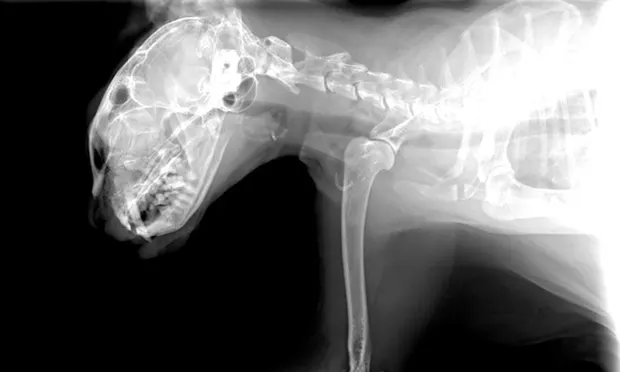
As in the majority of trauma cases, radiographs of the patient’s thorax and abdomen may be indicated. Skull radiographs provide little information regarding the severity of brain injury but may reveal structural damage such as fractures, especially if a physical defect in the skull is noted during physical examination. Testing such as computed tomography and magnetic resonance imaging may be required to further investigate the severity of brain injury.
Treatment Recommendations
Extracranial therapy includes maintenance of adequate intravascular volume, oxygenation, and ventilation. Avoidance of hypoxia and hypotension is vital to maximizing the chance of a successful outcome. Isotonic crystalloids should be administered at 10 to 20 mL/kg given over 10 to 15 minutes. Blood products should be considered in animals with significant hemorrhage. Hypertonic saline, a hypertonic crystalloid, is an excellent choice for resuscitation in TBI patients. The recommended dose is 4 to 6 mL/kg of 7.5% NaCl over 10 to 15 minutes. Oxygen therapy should be administered via flow-by mask, oxygen tent, or oxygen cage to maintain a pulse oximetery oxygen saturation >95%. Nasal oxygen tubes or cannulas should be avoided as these may cause sneezing and/or coughing, which can lead to ICP elevation.
Intracranial therapy involves minimizing increases in ICP and maximizing CPP. Increases in ICP can be minimized by limiting obstruction to venous drainage. Similarly, placing jugular central lines and occluding the jugular vein should be avoided. The patient’s head should be elevated at a 15°- to 30°-angle. When elevated ICP is suspected based on the presence of the Cushing reflex and acute deterioration of the patient’s status, hyperosmolar therapy should be initiated. Mannitol is given at a dose of 0.5 to 2 g/kg as a slow bolus over 15 to 20 minutes and should be administered through a filter to account for the possibility of crystal formation at lower temperatures and in concentrations >15%.9
Additional Therapies
Anticonvulsant therapy should be initiated as soon as a patient with TBI has a seizure. Analgesia is paramount in these patients, as it may help prevent further ICP elevation. Opioids are used commonly because they are easy to reverse and do not result in cardiac effects. The use of corticosteroids in veterinary head trauma patients is not currently recommended, as they have been shown in human clinical trials to increase mortality. Corticosteroid use in human head trauma patients has also been associated with hyperglycemia, immunosuppression, delayed wound healing, and gastric ulceration.1
Nutrition should be considered early, and electrolytes should be monitored often and supplemented as required. Special attention should be placed on serum glucose concentrations. Insulin therapy may be provided in animals that are persistently hyperglycemic. Other aspects of patient care include bladder management, frequent turning of the recumbent patient, and passive range-of-motion exercises.1
Prognosis
Few data are available regarding prognosis in animals with TBI. Potential complications include seizures, blindness, sepsis, and coagulopathies. Animals have a remarkable ability to compensate for loss of cerebral tissue. Neurologic recovery can encompass a variety of forms and may be complete, partial, or unacceptable to the owner of the pet. Some patients may lose housetraining ability and need to relearn the behavior, whereas other animals may return to normal function. It is impossible to provide a definitive prognosis on the degree of function that each individual patient will attain.
The initial appearance of a patient can be deceiving. Some animals may demonstrate mild clinical signs but rapidly deteriorate over the following 24 to 48 hours, whereas others may be severely affected at presentation but improve a great deal during this key period. The initial stages of care are critical and the patient’s response over this time may be an important indicator of prognosis. Appropriate therapy can significantly improve the outcome of a patient with TBI.