Trauma in a Mallard Hen
A 1.5-kg adult mallard hen was attacked by a raccoon approximately 4 hours prior to presentation.
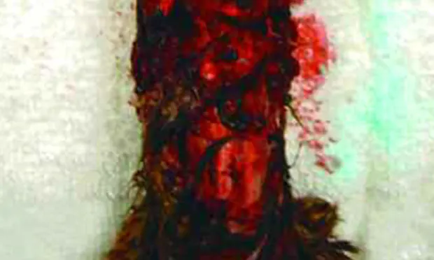
The owner reported the duck had severe blood loss immediately after the attack. The duck was quiet and depressed but responsive upon physical examination. There was an approximately 4 cm × 8 cm degloving injury to the dorsal cranial aspect of the neck region extending from the mid dorsal cervical area to the base of the skull (Figure 1).

The wound was not bleeding. The duck's heart rate (185 beats per minute) and respiratory rate (11 breaths per minute) were within normal limits (reference intervals: 175 to 194 and 8.2 to 12.6 per minute, respectively).1 A blood sample was obtained for a complete blood count (Table), and a swab of the wound was submitted for aerobic culture and sensitivity testing.

Ask yourself ...• What would you do to stabilize this critical avian patient?• How would you treat the injury?• What analgesics would you use?• What concerns do you have regarding the raccoon bite and what would you do to alleviate these concerns?• What is your diagnosis based on the initial history and physical examination?
Did You Answer ...• Initial assessment is required to determine how much stress, through handling, the patient can endure. Fluids to improve hydration and tissue perfusion, pain control, and prevention of bacterial septicemia are the main focus of initial treatment. Maintaining normothermia is extremely important. As soon as possible, the patient should be placed in an intensive care unit with oxygen support capabilities, digital temperature monitors, and reliable temperature control.
• The wound should first be cultured for bacterial isolates then aggressively cleaned and bandaged. There are 2 treatment options: One is second intention healing as was instituted in this case. The other option is secondary closure, managing the affected area until there is a granulation bed and the threat of infection has been eliminated; then using an advancement skin graft to close the area by primary intention healing.
• Meloxicam, a nonsteroidal antiinflammatory agent that can be administered orally and is very palatable, and butorphanol, a partial opiate agonist/antagonist, are appropriate.
• A major concern with a raccoon or cat bite is exposure to many pathogenic organisms. Often, the initial supportive care improves the patient's condition, only to have septicemia occur 72 to 96 hours later. It is therefore imperative that aggressive antibiotic therapy be initiated against both aerobic and anaerobic organisms.
• The history of trauma by a raccoon attack necessitates exploring the potential for internal injuries. In this case, endoscopic examination of the cranial gastrointestinal tract revealed an esophageal tear and proventricular granulomas, the latter of which were incidental findings.
Diagnosis
Severe degloving injury with potential for life-threatening septicemia
As with most trauma-induced injuries, one must consider both internal injuries and an affected area greater than that directly visualized. The extensive wound on the head reinforced the severity of the attack and potential for internal injuries.
Stabilization. The critical status of the duck focused initial medical attention on stabilization through fluid loss replacement, pain control, and prevention of bacterial septicemia. Normosol (25 ml/kg IV bolus over 5 min) delivered through a catheter placed in the medial tarso-metatarsal vein, 2 analgesic medications (butorphanol, 2 mg/kg Q 12 H IM and meloxicam, 0.2 mg/kg Q 12 H PO), and antimicrobial agents (piperacillin-tazobactam, 125 mg/kg Q 12 H IV and metronidazole, 15 mg/kg Q 12 H PO) were administered.
A swab was taken of the dorsal cervical wound for aerobic bacterial culture, which produced a mixed bacterial growth. The microbiologists determined the isolates as multiple contaminant organisms not worthy of further identification, and the lesion was cleaned, debrided, and flushed with saline and a 0.05% chlorhexidine solution. The degloving injury was extensive and could not be covered with remaining viable skin. The bite wound was left open for treatment and bandage changes. The duck was placed in a temperature-controlled incubator at 85°F, and fluid therapy (continuous rate, 100 ml/kg Q 24 H), and tube-feeding (Avian Critical Care; Lafeber Company, www.lafeber.com) were administered 2 to 3 times a day for the first 2 weeks.
Further Examination. After a few days of critical care and aggressive wound management consisting of dilute chlorhexidine flushes, debridement of nonviable tissue, antimicrobial therapy, and frequent bandage changes, the duck was stabilized. It was then anesthetized and an endoscopic examination was performed of the esophagus and proventriculus. Anesthesia was induced (5% isoflurane and 1 L/min oxygen) using a face mask and the patient was intubated and maintained on 2% isoflurane and 1 L/min oxygen. A small tear and numerous very small proventricular granulomas were observed in the distal esophagus. At the time, we believed the granulomas were incidental findings and not related to the predatory attack. Because the tear was small and due to its location, medical management was considered the best option. There was no gross evidence of trauma to the trachea or subcutaneous emphysema.
Follow-up & Outcome. During the first weeks of follow-up treatment, the external wound was flushed with chlorhexidine 0.05% before applying a layered bandage consisting of a hypertonic sponge (Curasalt; Tyco Healthcare, www.tycohealthcare.com) followed by an antimicrobial sponge (Kerlix AMD; Kendall Wound Care Products, www.kendallamd.com) and bandage material (cotton roll and Vetrap; 3M, www.3m.com). The bandages were changed daily. Surgical debridement and reconstruction of the wound were necessary every 3 to 4 days to protect as much of the remaining tissue as possible.

Once debridement of the wound was accomplished, the formation of granulation tissue was stimulated through application of hydrocolloidal gel and hydrogel wound dressing (Curagel; Tyco Healthcare, www.tycohealthcare.com) followed by bandage material (cotton roll and Vetrap). The bandages were changed every 3 to 4 days. When sufficient granulation tissue had formed (2 weeks after presentation), the wound was managed with a semiocclusive, hydrophilic foam dressing (Hydrasorb; Kendall Wound Care Products, www.kendallamd.com) in an attempt to stimulate epithelization. The wound eventually healed and the feathers regrew (Figures 2, 3A, and 3B).
Endoscopy was used to reexamine the esophageal tear; the affected area had healed. The incidental granulomas noted during the first examination were still present. Reexamination of the duck was scheduled 4 weeks after discharge, and confirmed resolution of the wound.
Bite wounds, especially from a cat or raccoon, present many pathogenic bacteria. In a survey of oral aerobic microbial flora of the raccoon, the average number of isolates per animal was 5, with a range of 3 to 9.2 The following aerobic bacterial isolates were identified in the oral cavity of raccoons:2 alpha, beta, and nonhemolytic Streptococcus, Staphylococcus, Bacillus, Micrococcus, Corynebacterium pyogenes, Citrobacter, Edwardsiella, Enterobacter, smooth and rough Escherichia coli, Klebsiella pneumoniae, Proteus, Providencia, Actinobacter, Aeromonas, and Pseudomonas. In addition to repairing any overt injuries, a quick response (aggressive wound care and antibiotic therapy) is necessary to check the potential for life-threatening septicemia.
TRAUMA IN A MALLARD DUCK • Thomas N. Tully, Jr and David Sanchez-Migallon Guzman
References1. Hematological and blood chemical values of mallard, Anas p. platyrhynchos, drakes before, during and after remige moult. Driver EA. J Wildlife Dis 17:413-421, 1981.2. Survey of the oral aerobic microbial flora of the raccoon. Mitchell MA, Hungerford LL, Nixon CM, et al. The NWRA Quart J, Summer:7-9, 1999.