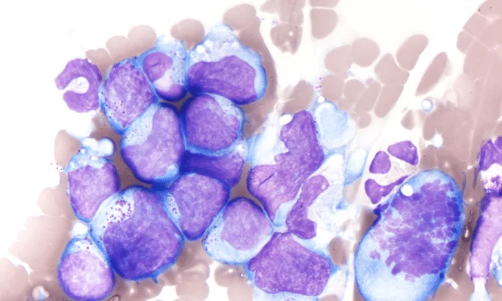
In the Literature
Parsley AL, Schnelle AN, Gruber EJ, Sander WE, Barger AM. Total protein concentration as a predictor of neoplastic peritoneal and pleural effusions of dogs. Vet Clin Pathol. 2022;51(3):391-397. doi:10.1111/vcp.13122
The Research …
Distinguishing neoplastic from nonneoplastic effusion in the pleural or peritoneal cavity can be a diagnostic challenge, particularly in samples with low cellularity. Biochemical parameters (eg, pH, lactate, glucose, vascular endothelial growth factor, telomerase) have been evaluated as markers of neoplastic effusion1-5; one study demonstrated higher lactate and lower glucose levels in neoplastic effusions compared with nonneoplastic effusions.2
Based on anecdotal observations that neoplastic effusions have higher total protein (TP) concentrations than nonneoplastic effusions, this study evaluated whether TP could be used to support diagnosis of neoplasia in effusion samples with lower cellularity (≤5,000 nucleated cells/µL). Medical records of dogs presented to a veterinary teaching hospital for nonhemorrhagic pleural or peritoneal effusion with ≤5,000 nucleated cells/µL were retrospectively reviewed. Cases were categorized as neoplastic or nonneoplastic, and the nonneoplastic group comprised 5 subcategories based on pathophysiologic cause of effusion (ie, decreased oncotic pressure, increased hydrostatic pressure, increased vascular permeability, leakage of urine, leakage of lymph). Fluid analysis, including TP, total nucleated cell count, erythrocyte concentration, specific gravity, and cytologic analysis, was performed on each sample. Serum albumin level and peripheral blood albumin:fluid TP ratio were also recorded.
Ninety-two cases met inclusion criteria; 27 had effusions that were neoplastic in origin. Pleural and peritoneal effusions in the neoplastic group had significantly higher TP levels compared with the nonneoplastic group; however, statistical significance was lost when the decreased oncotic pressure subcategory (n = 23) was excluded from the nonneoplastic group. The neoplastic group also had a significantly lower peripheral blood albumin:fluid TP ratio than the nonneoplastic group. Effusions with a ratio ≤0.6 were 5.6 times more likely to be neoplastic; however, most of those cases had infiltrative primary or metastatic liver neoplasia, which may have influenced findings.
The authors concluded that evaluating fluid TP alone is unlikely to aid in distinction between neoplastic and nonneoplastic effusion in dogs because the difference in TP concentration between the study groups was primarily due to hypoalbuminemia.
… The Takeaways
Key pearls to put into practice:
Identifying the cause of effusion with low cellularity can be challenging. Additional point-of-care diagnostics and reliable parameters are needed.
TP values of effusions are unlikely to help distinguish between neoplastic and nonneoplastic causes, especially when used alone.
Using a combination of clinical findings, diagnostic parameters, and molecular diagnostics (eg, PCR for antigen receptor rearrangements, flow cytometry) when appropriate may increase confidence when identifying neoplastic effusions.
You are reading 2-Minute Takeaways, a research summary resource presented by Clinician’s Brief. Clinician’s Brief does not conduct primary research.