Differentiation between syncope and seizures is essential for successful treatment of underlying disease. Loss of consciousness is the most notable sign for both syncope and seizures.1,2 Clinical signs of syncope may also include collapse after specific activities (eg, standing, walking, running, coughing, barking), positional change, and occasional loss of bladder or bowel function. Clinical signs of seizures may also include somatic movement (eg, twitching of the facial muscles, limbs, or tongue) and autonomic activation (eg, urination, defecation, salivation, pupillary dilation), which can be subtle.3 Distinction between syncope and seizures (Table) is needed before diagnostic testing or treatment options are initiated because the pathophysiology, differential diagnoses, diagnostic testing, and treatment plans are markedly different.
Table: Syncope vs. Seizures Findings in Dogs and Cats
An objective scoring system for differentiating between syncope and seizures has been developed in human medicine.4 When associated with loss of consciousness, the scoring system identifies the following signs as highly indicative of a seizure in humans: evidence of a cut tongue, postevent confusion, head turn during unconsciousness, and some behavior or sensory signs that are difficult to identify in veterinary patients. The following signs are associated with a syncopal event: collapse without loss of consciousness, loss of consciousness with prolonged sitting or standing, and some behavior or sensory signs that are difficult to determine in veterinary patients. Although similar data are not available in veterinary studies, many of these signs may be transferable to veterinary patients due to a shared pathophysiology between species.
Following are the author’s top 5 methods for distinguishing syncope and seizures in dogs and cats.
1. Historic Evidence of Activity or Sleep
Syncope occurs secondary to cardiovascular events (eg, arrhythmia, hypotension, hypertension) or to changes in thoracic or abdominal pressure (eg, coughing, defecation). Prolonged standing, prolonged sitting, and rapid transition from lying to sitting, standing, or walking (ie, orthostatic hypotension) can result in a syncopal event in predisposed patients. It is important to question pet owners about any activity that may have occurred immediately prior to loss of consciousness. Patients with syncope commonly have a history of moving or standing when unconsciousness occurs. Conversely, seizures occur more commonly when a patient is at rest rather than in motion.
Whether exercise is neuroprotective for dogs and cats remains unknown, but some evidence supports this phenomenon in humans with epilepsy.5-7 Although activity may reduce the incidence of seizures, seizure during activity is still possible; therefore, all aspects of the history and physical examination should be considered when determining whether the patient experienced a seizure or syncopal event.
2. Autonomic Signs
Loss of bladder control has been reported in patients with syncope and is common in patients with seizures.2 Other autonomic changes (eg, loss of bowel function, salivation, lacrimation, pupillary dilation) have not been reported in patients with syncope but are regularly reported in patients with seizure disorders.8 Activation of autonomic networks in the brain are responsible for seizure-induced autonomic signs9; therefore, consistent autonomic signs suggest an epileptic disorder rather than a syncopal cause.
3. Behavior Changes
Syncope
Syncope is caused by transient loss of blood flow to higher brain centers from a cardiovascular origin; therefore, neuronal resetting is not needed when blood flow is restored, and return to normal mentation is rapid. Patients with syncope commonly recover rapidly and regain complete consciousness without behavior changes.
Preictal Phase of Seizures
Behavior changes (eg, pacing, seeking out owners, hiding) may be present prior to seizure onset and may suggest a preictal state. Preictal signs are not identified in all patients with every ictus; therefore, this is a less reliable indication of an impending seizure.
Postictal Phase of Seizures
Evidence of confusion or changes in behavior following loss of consciousness are key indicators of a seizure disorder in humans and veterinary patients. Patients with seizures may exhibit pacing, anxiety, aggression, confusion, and compulsive movement when recovering from a seizure.3 Seizures are caused by hypersynchronous neuronal activity that results from an imbalance of excitation and inhibition in the brain. Recovery requires rebalancing excitation and inhibition, resulting in a prolonged, but transient, change in behavior.
4. Abnormal Physical or Neurologic Examination Findings
Complete physical and neurologic examinations are imperative when evaluating patients with a history of transient unconsciousness.
Syncope is cardiovascular in origin. Evidence of cardiac arrhythmia, cardiac murmur, asynchronous or weak pulse, or signs of poor perfusion (eg, blue or white mucous membranes) suggest cardiovascular disease should be further investigated.
Seizures originate from the prosencephalon (ie, forebrain). Identifying neurologic examination abnormalities referrable to forebrain dysfunction supports evidence of a seizure disorder. These findings include unilateral or bilateral menace deficits with intact pupillary light and dazzle reflexes (Figure 1), unidirectional compulsive circling without head tilt, unilateral sensory deficits to the face (hypoanalgesia or analgesia), changes in mental status, unilateral postural reaction deficits (Figure 2), and, in rare situations, evidence of hemiparesis.

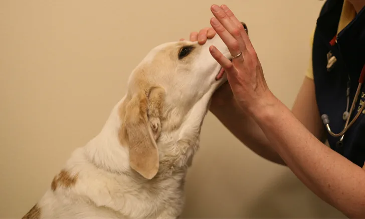
Abnormal menace response, especially if unilateral, with an intact pupillary light and dazzle reflex suggests dysfunction of the forebrain and, thus, a seizure disorder. Patients with syncope have a normal neurologic examination. The menace response is performed by advancing a hand briskly toward the patient’s eye and watching for a blink reflex. During an abnormal response, the patient does not blink; however, touching the medial or lateral canthus induces a blink reflex.

Dog with a paw replacement deficit noted on neurologic examination. This occurs more often with seizures because the forebrain is affected. Patients with syncope demonstrate a normal neurologic examination unless concurrent neurologic disease is present.
5. Evidence of Metabolic Disease
Following complete physical and neurologic examination, clinical pathologic evaluation can help identify possible metabolic causes of syncope or seizures. Cardiac troponin I is a myocardial-specific protein found in the contractile apparatus of the heart.11 Increased serum cardiac troponin I levels have been measured in humans with seizures and syncope and were found to be significantly more elevated in patients with syncope than in those with seizures; however, severe seizure disorders (eg, status epilepticus) can cause myocardial damage and may also result in elevated serum cardiac troponin I levels. In one veterinary study, serum cardiac troponin I levels were found to be significantly different between dogs with seizures and dogs with syncope11; however, notable overlap was found, thus rendering the test ineffective for definitively differentiating between the diseases in dogs. Troponin I levels do not reflect all causes of syncope.
Metabolic abnormalities associated with seizures, including hypoglycemia, hypocalcemia, elevated ammonia, and hypernatremia, can be readily identified on routine serum chemistry analysis. Evidence of these metabolic disorders concurrently identified in a patient with a history of acute loss of consciousness should lead to consideration for seizure disorder. Patients with metabolic disease can have concurrent cardiovascular disease; inclusion of other differentiating factors should be considered.
In human medicine, serum lactate levels measured within 2 hours of the episode of lost consciousness are significantly higher in patients with seizures compared with patients with syncope.10 This has yet to be verified in veterinary medicine but may become a readily available test to objectively differentiate between seizures and syncope in veterinary patients.
Conclusion
Syncope and seizures are caused by different pathology, but both conditions can result in an acute, transient loss of consciousness. Thorough patient history, physical examination, neurologic examination, and serum chemistry assessment can improve the ability to differentiate between these diseases and improve long-term patient care.